Playlist
Show Playlist
Hide Playlist
Anatomy of the Diaphragm

-
Slides Anatomy of the Diaphragm.pdf
-
Download Lecture Overview
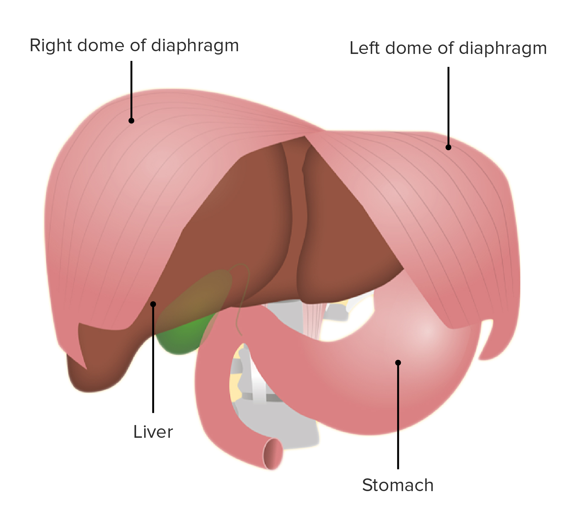
00:01 Now we're going to talk about one of the most important muscles in the entire body, the diaphragm. 00:06 It's so important because the diaphragm is the muscle that primarily helps us breathe. 00:11 It's also what separates the thorax from the abdomen. 00:15 In fact, phragm means fence. 00:18 So it's essentially the fence that separates the thorax from its next door neighbor, the abdomen. 00:24 The diaphragm is kind of a complicated shape, and there's a lot going on, so use yours to take a deep breath, and we'll dive right in. 00:33 The diaphragm occupies and seals off essentially, the inferior thoracic aperture. 00:40 Therefore, all of its attachments are going to mirror the borders of the inferior thoracic aperture. 00:46 For example, we'll have this xiphoid part where it's joined to the xiphoid process. 00:52 The costal part where it's joined to ribs seven through 12. 00:57 And the lumbar part, downward attaches to the lumbar vertebra L1 to L3. 01:04 If we look from above, if we have a superior view, looking down on the diaphragm, we see that this sort of dome shaped muscle has fibers that travel centrally. 01:16 And they meet up at this large flat sheet, sometimes called an aproneurosis, but specifically here called the central tendon. 01:24 And so as these fibers contract, it will draw the diaphragm inferiorly, which will increase the thoracic cavity volume, which will lower the pressure, and will subsequently cause the lungs to expand. 01:39 And so that's how the diaphragm helps us breathe. 01:44 Now, it's great that it fills the entire inferior thoracic aperture. 01:49 But there are some structures that still need to pass between the thorax and the abdomen. 01:53 So we have some openings. 01:55 We see one right here in the central tendon called the caval opening. 02:00 And it's called that because that's where the inferior vena cava is going to pass through to reach the right atrium. 02:07 Just to the left and posterior to that we have another opening and that's the esophageal hiatus, where the esophagus is going to pass through on its way to the stomach. 02:17 And then most posteriorly we have this hole here, which is the aortic hiatus. 02:22 Where the thoracic aorta is going to pass through and then on the other side turn into the abdominal aorta. 02:29 Now let's look at those openings from below. 02:32 So if we look from below, from the abdomens point of view, we again see the caval opening. 02:39 And for reference, to give you an idea, roughly of where we're at, we're at about the T8 vertebral level. 02:45 And as the name implies, the inferior vena cava is going to pass through there. 02:51 We're also going to have some branches of the right phrenic nerve taking advantage of this opening. 02:56 Just to the left and posterior is that other opening about the T10 level. That's the esophageal hiatus. 03:04 Where the esophagus is going to pass through. 03:07 And we're also going to have some trunks of the vagus nerve. 03:11 Then finally, most posteriorly we have that aortic hiatus all the way down at around the T12 level, where the aorta is going to transition from being a thoracic aorta to an abdominal aorta. 03:24 And it's also where the thoracic duct is going to be passing from the admin up into the thorax. 03:32 If we focus a little bit more on the inferior and posterior portions of the diaphragm, we see some weird stuff going on. 03:39 We have what are called crura. Crura just means the legs. 03:42 And we have a right leg or a right crus and a left leg or a left crus. 03:48 On either side of the inferior most portion of the diaphragm here. 03:54 And if we zoom in in that area, and we look at the area of the diaphragm, especially around the esophageal hiatus where the esophagus is coming through, we see it kind of divides. 04:06 We see some of that dividing into one leg off to the right, the right crus, and then the other off to the left, which called the left crus. 04:15 There's this weird little thing happening off of the right crus. 04:18 We see some of it going over and around and past the celiac trunk of the abdominal aorta, on its way to a portion of the intestine called the duodenum. 04:30 And that's why this funny little muscle is called the suspensory muscle of the duodenum. 04:35 Or you may have heard it by its eponym the ligament of Treitz. 04:39 If you've had embryology, you've probably heard of this as an important landmark for rotation of the gut. 04:44 But it's also an important landmark in just anatomic sense because this is what we generally say divides the GI tract into upper and lower. 04:54 So for example, if you're saying an upper GI bleed versus a lower GI bleed, this is to the point where we're talking about upper becomes lower. 05:04 Alright, so let's talk about the innervation a little bit. 05:07 So, we have the phrenic nerves, and the phrenic nerves originate up in the neck and traveled down through the mediastinum on their way to the diaphragm to provide motor innervation there, but we also see as it's going through the mediastinum. 05:23 it's giving off branches. 05:24 And we see for example, there's some pericardial branches here. 05:29 And so there's going to be a lot of sensory innervation provided by the phrenic nerves before they reach the diaphragm to provide motor innervation. 05:39 And they're going to go all the way to the undersurface or the abdominal surface, where they're going to terminate as phrenicoabdominal branches very appropriately named. 05:52 Sometimes there can even be a little ganglion called the phrenic ganglion that will communicate with the celiac ganglion down in the abdomen.
About the Lecture
The lecture Anatomy of the Diaphragm by Darren Salmi, MD, MS is from the course Thorax Anatomy.
Included Quiz Questions
What is the most inferior attachment of the diaphragm to the spinal column?
- L3
- L1
- S1
- T12
- S3
What is the spinal level of the caval opening?
- T8
- T10
- T12
- L2
- L4
Which group of neurons may communicate with the phrenic nerve and the phrenic ganglion?
- Celiac ganglion
- Superior mesenteric ganglion
- Inferior mesenteric ganglion
- Sacral plexus
- Epigastric plexus
Author of lecture Anatomy of the Diaphragm

Darren Salmi, MD, MS
Customer reviews
5,0 of 5 stars
| 5 Stars |
|
5 |
| 4 Stars |
|
0 |
| 3 Stars |
|
0 |
| 2 Stars |
|
0 |
| 1 Star |
|
0 |
