Playlist
Show Playlist
Hide Playlist
Visual Pathway

-
Slides Anatomy Visual Pathway.pdf
-
Download Lecture Overview
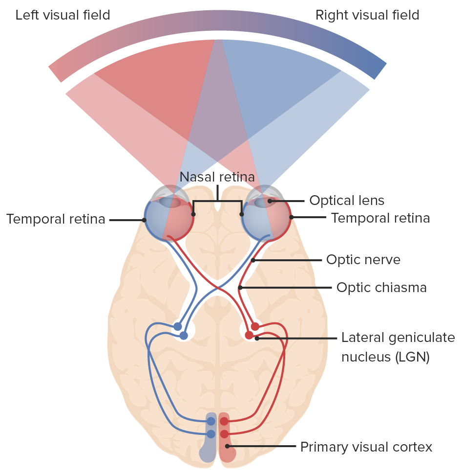
00:02 Now let's take a closer look at the optic nerve, cranial nerve II. 00:07 Here we see the sphenoid bone and its relationship to the optic nerve coming from the eyeball. 00:14 As it moves posteriorly, it's going to pass through the opening of the sphenoid bone known as the optic canal. 00:20 Before it interacts with its contralateral side at the optic chiasm. 00:25 Before continuing on more posteriorly into the brain as the optic tract. 00:31 When we look at the retina, which is the sensory apparatus of vision, we can divide it into medial and lateral parts. 00:38 We also call it the nasal and temporal parts because the medial part is closer to the nose and the temporal part is closer to the temporal area of the skull. 00:48 It turns out, when we're looking at a particular area or a visual field, the temporal aspect of the visual field is actually received by the nasal aspect of the retina and vice versa. 01:03 The temporal aspect of a retina is picking up the nasal aspects of the visual field. 01:09 As you can see by the color coding in this image here. 01:13 Furthermore, the fibers that carry those visual field informations travel in different aspects, so the temporal fibers, again those are carrying information from the nasal fields stay somewhat laterally, whereas the nasal fibers, seeing somewhat more medially, will actually cross over at the optic chiasm. 01:39 And this is clinically important because this area of the optic chiasm happens to be right near the pituitary gland. 01:46 So, when the pituitary gland becomes abnormally enlarged, such as with a pituitary adenoma, it may compress this optic chiasm. 01:56 And again, this is where those nasal fibers are crossing. 02:00 And again, keeping in mind that nasal fibers carry the temporal fields, leading to a particular type of vision loss called bitemporal hemianopsia. 02:12 In fact, when you see this type of visual field loss, it helps you target a potential pathology related to the pituitary gland. 02:25 Now let's take a look at how the orbit interacts with the middle cranial fossa. 02:31 Here we see the optic nerve reaching the optic chiasm or that crossing point. 02:38 Beyond which, it becomes the optic tracts as they go to the visual processing areas. 02:44 And the optic canal is a passageway through the sphenoid bone. 02:50 The superior orbital fissure, also a feature of the sphenoid bone carries multiple cranial nerves: III, IV, V1 and VI. 03:00 One thing that does not carry is the maxillary division of trigeminal that goes through the foramen rotundum.
About the Lecture
The lecture Visual Pathway by Darren Salmi, MD, MS is from the course Special Senses.
Included Quiz Questions
The optic canal passes through which bone?
- Sphenoid
- Nasal
- Frontal
- Ethmoid
- Maxilla
What pathology is associated with bitemporal hemianopsia?
- Compression of the optic chiasm
- Retinal scarring
- Lesion to the optic nerve
- Lesion to the optic tract
- Lesion to temporal fibers
Author of lecture Visual Pathway

Darren Salmi, MD, MS
Customer reviews
5,0 of 5 stars
| 5 Stars |
|
5 |
| 4 Stars |
|
0 |
| 3 Stars |
|
0 |
| 2 Stars |
|
0 |
| 1 Star |
|
0 |
