Playlist
Show Playlist
Hide Playlist
Cervical Vertebrae and Hyoid Bone

-
Slides Anatomy Cervical Vertebrae Hyoid Bone.pdf
-
Download Lecture Overview
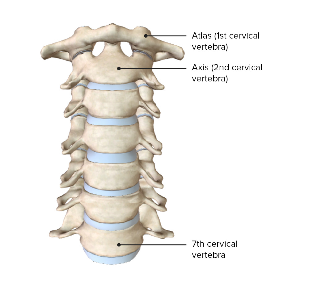
00:01 Let's begin our discussion of neck anatomy by looking at the bony anatomy of the neck, which is mostly the cervical vertebrae, as well as one special bone called the hyoid. 00:13 Here we see the cervical vertebrae, which we number 1 to 7 from superior to inferior. 00:19 We also give the first two cervical vertebrae, unique names because of their unique shape and function. 00:26 C1 is also called atlas and C2 is also called axis. 00:30 We then have C3, 4, 5, 6. 00:33 And finally C7, which also gets a special name vertebra prominens because it projects very prominently posterior more than any of the other vertebra, and makes it a useful landmark during physical examination. 00:48 Here's a typical cervical vertebrae. 00:50 We'll look at the typical ones first, and then examine the differences between these and C1 and C2. 00:58 We see that the body is somewhat square shaped here. 01:02 The superior surface of the body is a bit concave while the inferior surface is a bit convex. 01:10 Here we have a superior view where we can see the body and we see the spinous process tends to be bifid or split. 01:18 And that's usually in C3 to C6. 01:22 We have the lamina that connects it to the rest of the vertebral arch and the pedicle that connects it to the body. 01:29 Here the vertebral foramen where the spinal cord travels is a bit triangular. 01:36 On this lateral view, we see the inferior and superior articular process or facet. 01:43 We also see in the transverse process, an opening, something we don't see in the rest of the vertebrae. 01:49 And this is the transverse foramen. 01:53 And this is an important landmark that is unique to the cervical vertebrae. 01:57 because these transverse foramina collectively are where the vertebral artery passes through. 02:04 The vertebral artery is real important artery because it's going to form anastomosis in the brain called the circle of Willis. 02:11 Usually it does skip the C7, although on some occasions it will go through that foramen as well. 02:21 Now here are the atypical vertebrae that look pretty different from the others. 02:24 Starting with C1 or the atlas. 02:28 We don't really see much of a body here instead we have this bump called the anterior tubercle on the anterior arch, which connects to these lateral masses. 02:38 They do have a transverse process and a transverse foramen. 02:42 But instead of a superior articular facet, we have a facet for the occipital condyle, which is a bump on the occipital bone. 02:51 And then posteriorly we have a posterior arch with a posterior tubercle. 02:58 C2 is also pretty atypical. 03:01 Here we see the articular surface for atlas or C1 upon which it's going to rest. 03:06 And the unique feature here is this projection called the dens or sometimes odontoid process. 03:15 And when we put these two vertebrae together, we have the atlantoaxial joint. 03:20 And now we can see that C1 or atlas rotates around this dens and it allows us to have this rotating action as if we were shaking our head no. 03:33 The atlanto-occipital joint, on the other hand is going to connect the vertebrae to the skull. 03:40 That's why C1 gets the name atlas. 03:43 It's sort of like the Greek figure that's holding the globe on the shoulders. 03:47 Here we see C1. And here are some condyles on the occipital bone. 03:52 They're going to articulate with it. 03:55 As they come together, this allows for a flexion, extension motion between C1 and the skull. 04:03 Much like nodding your head yes. 04:08 Finally, let's look at the hyoid bone. 04:11 The hyoid is this U shaped bone that's composed of a body as well as a greater horn and a smaller more anteriorly placed lesser horn and many important ligaments and muscles are going to attach to this bone and are going to be very important for the act of swallowing.
About the Lecture
The lecture Cervical Vertebrae and Hyoid Bone by Darren Salmi, MD, MS is from the course Neck Anatomy.
Included Quiz Questions
What is another name for the C1 vertebra?
- Atlas
- Axis
- Dens
- Hyoid
- Larynx
What is the shape of a typical cervical vertebral body?
- Square
- Circular
- Rectangle
- Triangular
- Hexagon
What is a unique feature of the cervical vertebrae?
- Transverse foramen
- Superior articular facet
- Inferior articular facet
- Lamina
- Pedicle
Author of lecture Cervical Vertebrae and Hyoid Bone

Darren Salmi, MD, MS
Customer reviews
5,0 of 5 stars
| 5 Stars |
|
5 |
| 4 Stars |
|
0 |
| 3 Stars |
|
0 |
| 2 Stars |
|
0 |
| 1 Star |
|
0 |
