Playlist
Show Playlist
Hide Playlist
Bony Orbit
-
Slides Anatomy Bony Orbit.pdf
-
Download Lecture Overview
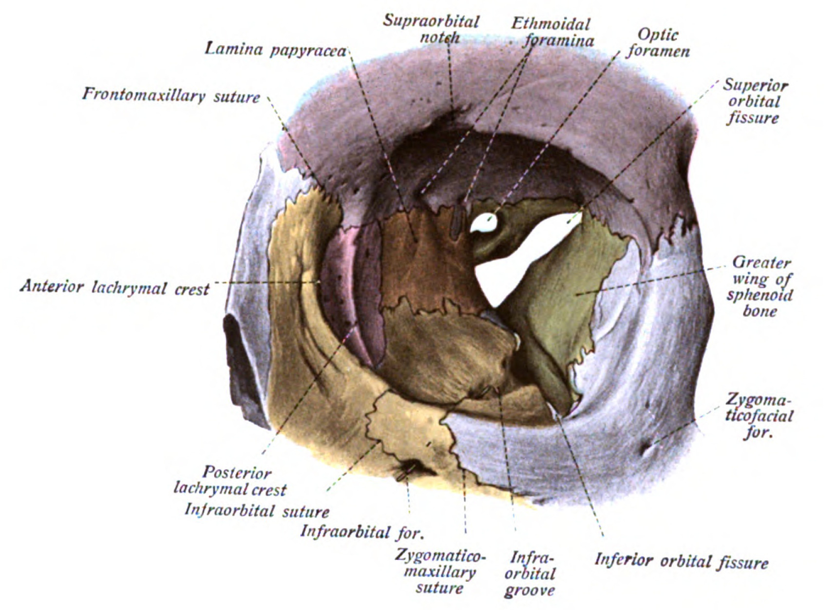
00:01 Now we're going to talk about the orbit. 00:04 The orbit is a very special cavity within the skull where we're going to find the eyes. 00:11 The orbits are gonna be formed by many different bones. 00:15 They're going to form a space for other structures to fill in. 00:20 So after we talk about the bones that outline the orbit, we're going to start filling in some of those structures. 00:25 Most importantly, we're going to talk about the eyeball. 00:29 We're also going to talk about the stuff that's found inside the eyeball, things like the retina, the lens and the iris. 00:37 We're also going to zoom out a little bit and look at the muscles in the orbit that act upon the eyeball to move it around and direct where eyes are looking. 00:47 Then we're also going to look at something called the lacrimal system, which bays are eyes in tear fluid to keep them from drying out naturally help us with our vision. 00:59 The other things is going to protect our eyes or the eyelids. 01:02 So we're also going to cover those and talk about some of the external and internal features of the eyelids as well. 01:09 Now let's starting with the bony orbit. 01:13 Here we see parts of the frontal bone, maxillary bone, ethmoid bone, sphenoid bone, lacrimal bone, and zygomatic bone, as well as a little bit of the palatine bone and they all contribute to the orbit. 01:32 The orbit itself is somewhat pyramidal shaped with a very wide opening or base anteriorly and getting narrow as it moves posteriorly towards the apex. 01:45 Therefore, it also has a roof, a floor, a medial wall, and a lateral wall. 01:54 If we take a closer look at the roof, we find the orbital plate of the frontal bone as well as the lesser wing of the sphenoid bone. 02:05 We see some features in this roof such as a little depression called the trochlear fovea, which is where we're going to find a little structure called the trochlea. 02:15 We also see a depression called the lacrimal fossa, which is we're going to find that lacrimal gland. 02:22 If we look over at the medial wall, here, we find the lacrimal bone and the orbital plate of the ethmoid bone as well as a bit of the body of the sphenoid. 02:33 We also find some openings here such as the anterior and posterior ethmoidal foramena. 02:39 As well as a fossa for something called the nasal lacrimal sac. 02:45 On the floor, we have the orbital plate of the maxilla as well as some of the zygomatic bone. 02:53 We also have the orbital process of the palatine bone. 02:58 We have another space here, this time called the inferior orbital fissure. 03:03 And we can see that that's going to lead to an opening called the infra orbital canal that opens up as the infraorbital foramen anteriorly. 03:14 With the lateral wall, we see more of the zygomatic bone as well as the greater wing of the sphenoid. 03:21 We have openings here as well such as the zygomaticotemporal and zygomaticofacial phenomena. 03:29 We also see the superior orbital fissure. 03:34 Now let's look at those openings again. 03:37 We have a little opening at the superior most aspect of the orbit called the supraorbital notch or foramen depending on how complete the hole is. 03:47 And this is where we're going to find passageways for our supraorbital neurovasculature. 03:53 We also see an opening called the optic canal through which the optic nerve or cranial nerve II is going to pass. 04:02 The much larger opening called the superior orbital fissure is going to contain multiple nerves such as cranial nerves III, IV, V1, and VI. 04:16 We also have a corresponding inferior orbital fissure. 04:20 That's we're going to find a branch of the trigeminal nerve called the maxillary nerve or cranial nerve V2. 04:27 We also see the infraorbital foramen again, and that's going to be the passageway for the infraorbital neurovasculature. 04:35 We see this nasolacrimal canal which is going to be where we find the nasolacrimal duct. 04:43 And the nasolacrimal duct, as the name implies, is gonna have something to do with tear drainage. 04:50 We find the ethmoidal foramena, the anterior and posterior ethmoidal arteries passing through those. 04:59 And with the zygomatic temporal foramen, we have the zygomatic temporal nerve and similarly the zygomaticofacial foramen with the zygomaticus facial nerve.
About the Lecture
The lecture Bony Orbit by Darren Salmi, MD, MS is from the course Special Senses.
Included Quiz Questions
Which bone is NOT part of the bony orbit?
- Nasal
- Frontal
- Sphenoid
- Zygomatic
- Lacrimal
What composes the roof of the bony orbit? Select all that apply.
- Orbital plate of the frontal bone
- Lesser wing of the sphenoid
- Lacrimal bone
- Orbital plate of the ethmoid bone
- Body of the sphenoid
Which bones compose the floor of the bony orbit? Select all that apply.
- Orbital plate of the maxilla
- Orbital process of palatine bone
- Zygomatic bone
- Greater wing of the sphenoid
- Lesser wing of the sphenoid
Customer reviews
5,0 of 5 stars
| 5 Stars |
|
5 |
| 4 Stars |
|
0 |
| 3 Stars |
|
0 |
| 2 Stars |
|
0 |
| 1 Star |
|
0 |


