Playlist
Show Playlist
Hide Playlist
Thoracic Skeleton

-
Slides Anatomy Thoracic Skeleton.pdf
-
Reference List Anatomy.pdf
-
Download Lecture Overview
00:01
The thorax is home to many
interesting and
important structures.
00:05
But before we can
talk about those structures,
we have to talk about the
bony elements that enclose them.
00:12
Particularly, we're going to talk
about the sternum and the ribcage.
00:16
The sternum is this bone found
right along the anterior midline
that's broken up into three parts.
00:24
We have a Manubrium.
We have a body.
00:29
And at the inferior edge,
we have a xiphoid process
or xiphi sternum.
00:35
And that xiphoid process you might
notice looks a little pointy.
00:38
That's because long ago when
people used to use swords,
it looked to them like the
sternum was an upside down sword.
00:45
In fact, xiphoid means sword
like and manubrium means handle.
00:50
Speaking of the manubrium,
there's a little feature
we want to point out
and it's that little divot
along the superior edge.
00:57
That's called the jugular
or suprasternal notch.
01:01
And that's a landmark that's
usually pretty easily palpated
during a physical exam.
01:07
These parts of the sternum
have joints with each other,
although they're not the
typical joints you might think of.
01:12
These are synarthrosis
meaning there's really
no movement between them.
01:16
But we have a joint between
the maneuver human body called
the manubriosternal joint
also called the sternal angle
or angle of Louis.
01:25
And then a joint between
the body and xiphoid process
called the xiphisternal joint.
01:30
And the fact that the
manubriosternal joint
has three total names is kind
of a clue that it's important.
01:35
And it is because it's the
anterior most projection
and therefore pretty easily
palpated all along the sternum,
and it's also where the
second rib attaches.
01:46
So it becomes a useful landmark
on physical exams.
01:49
The sternum also interacts with some
other bones, including the clavicle.
01:54
So the clavicle joins the manubrium
at the sternoclavicular or SC joint
and then the costal cartilages
are the cartilaginous ends
of the first seven ribs attached
to the sternum at something called
the sternalcostal or
sternalchondral joints.
02:14
Costal referring to rib.
02:16
Chondral referring to the
cartilaginous portion of the rib.
02:20
Speaking of ribs,
most people have 12 pairs,
and they're numbered from
superior to inferior 1 to 12.
02:30
And as we're already alluded to,
as they come around anteriorly
towards the sternum.
02:35
They turn from bone into cartilage,
and we call these the
costal cartilages.
02:41
And it's great that the ribcage
is very strong and protective
and protects the structures
that lie underneath it.
02:47
But there are still structures
that need to pass
in between the neck and the thorax.
02:53
And they do so through
an opening called
the superior thoracic aperture
or the thoracic inlet.
02:59
And this inlet aperture is
bordered by the T1 vertebra,
the edges of the first ribs,
and the manubrium.
03:09
Similarly,
there's an opening at the bottom
called the
inferior thoracic aperture.
03:16
And those borders
are the T12 vertebra, the 12th rib,
and those cartilaginous ends
of ribs 7 to 10.
03:26
And these xiphoid process
anteriorly.
03:31
And because these
cartilages all form
a pretty prominent inferior border,
sometimes we just refer to
this as the costal margin.
03:40
Talking a little bit about
the ribs in greater detail,
we sub classify them
as being true ribs
if they're the first
through seventh ribs,
because they attach
directly to the sternum.
03:52
And then we say ribs 8 through 12
are false because they don't.
03:57
Furthermore, we call ribs
11 and 12 floating ribs.
04:03
Now that's in contrast
to ribs 8, 9, and 10
that actually attach to each other
and then eventually
up to the seventh rib
via interchondral joints.
04:14
So they at least have an indirect
connection to the sternum.
04:18
So far, we've been focusing on
the anterior aspect of the ribs,
but they go all the
way around posteriorly
to meet up with the vertebra.
04:25
And the vertebra will be
covered in the back section.
04:28
But right now we're just going
to look at a transverse view
to where we can see the vertebral
body and the transverse processes.
04:37
The part of the rib that
interacts with the vertebral body
is the head of the rib.
04:42
And then the little bump
that interacts with
the transverse process
is called the tubercle of the rib.
04:48
And in between, we have something
called the neck of the rib.
04:52
If we go along a little bit
laterally from there,
we see that the rib
takes a sharp turn
and where it does so we call that
the angle of the rib
where these bones interact,
we have joints.
05:06
And between the tubercle of the rib
and the transverse process,
we have the Costotransverse joint.
05:12
And between the head of the rib
and the vertebral body,
we have the Costcovertebral joint.
05:17
And these are
true joints or synovial joints
that have a lot of movement.
05:22
But because there's
two of them here,
it provides an added layer
of stability to the ribs.
05:30
And the last part of
the rib we'll talk about
is this linear indentation along
the inferior interior edge,
called the costal groove.
05:38
And we'll see later when we talk
about the intercostal space,
that's where the
neurovascular structures
in this area
are going to run.
05:47
Now, for the most part,
ribs are just how we describe them,
and they're pretty similar
to each other.
05:52
But certain ribs have
some unique features
that are worth pointing out.
05:57
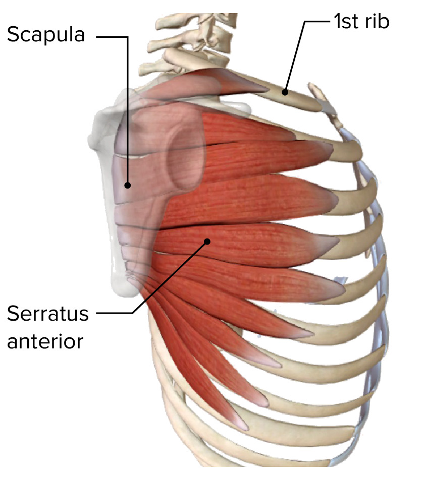
For example, the first rib
is really close to the neck.
06:00
So it's an ideal location to have
some attachments for neck muscles.
06:04
And in particular,
there's a little bump
called the scalene
tubercle on the first rib,
which is where the anterior
scalene muscle attaches.
06:13
And just anterior to that is a
groove for the subclavian vein,
because that's where the
subclavian vein runs,
and posterior li to that
muscles a little groove
for the subclavian artery
because that's where the
subclavian artery runs.
06:28
The second rib is
also pretty important,
because we already mentioned it
as a useful landmark
because it's where the
sternal angle is located.
06:37
And we can palpate that,
and it's good to know
where your second rib is
because usually can't
palpate your first rib
because it's usually
hidden by the clavicle.
06:47
And it's the sternal angle that
you're going to be looking for
when you're really trying
to number your ribs.
06:53
Really rib number two,
not rib number one.
06:57
The second rib still
close to the neck,
and it has attachment
points for neck muscles.
07:02
In this case, it has an attachment
for the posterior scalene.
07:05
Although, it's usually obscured
by the serratus anterior muscles.
07:11
The 12th rib is another weird one
because it only has a
single articular surface.
07:17
And it only articulates with
the vertebra at the head,
meaning it doesn't have
any other features like
a neck, a tubercle,
an angle or even a costal groove.
07:26
Now, 12 pairs is the
most common arrangement,
but people are going
to have more or less
or in fancier terms we say
supernumerary or infranumerary ribs.
07:38
For example,
about 1 in 200 people
have a rib that's coming
off of this C7 vertebra
up in the cervical area
called a cervical rib.
07:49
Usually it's bilateral meaning
there's one on both sides.
07:52
But it can be unilateral,
meaning just one on one side.
07:57
Most of the time, there's no
problems people are asymptomatic,
but it can cause
compression of structures
and cause something called
Thoracic Outlet Syndrome.
08:08
And that's because there's a
lot of important structures
that pass through this area,
including the brachial plexus.
08:15
So compression of the
brachial plexus can end up
causing pain or numbness
down in the forearm or hand.
08:22
And there's the subclavian
artery and vein in this area too.
08:26
So you can compress those vessels.
08:28
And that gets even worse
if the arm is abducted,
worsening that compression.
08:34
And in the case of compressing
the subclavian vein
that can limit how much blood
can return back to the body
causing the upper limb to swell up.
08:42
But again most of the time,
cervical ribs
cause no problem at all.
About the Lecture
The lecture Thoracic Skeleton by Darren Salmi, MD, MS is from the course Thorax Anatomy.
Included Quiz Questions
What is the most inferior bone of the sternum?
- Xiphoid process
- Body
- Manubrium
- Costochondral cartilage
- Clavicle
What is a synarthrosis?
- A joint that allows no movement.
- A joint that allows 180-degree rotation.
- A joint that allows 90-degree rotation.
- A joint in the lower half of the body.
- A joint in the upper half of the body.
Which rib attaches to the manubriosternal joint?
- 2nd rib
- 4th rib
- 5th rib
- 6th rib
- 8th rib
What differentiates the true and false ribs?
- The true ribs' costal cartilages articulate directly with the sternum.
- The true ribs' costal cartilages articulate indirectly with the sternum.
- The false ribs attach directly to the transverse process of the vertebral body.
- The true ribs attach directly to the transverse process of the vertebral body.
- The true ribs are formed prior to the false ribs during embryological development.
Which movement worsens symptoms of thoracic outlet syndrome?
- Limb abduction
- Limb adduction
- Limb flexion
- Limb extension
- Laying down
Author of lecture Thoracic Skeleton

Darren Salmi, MD, MS
Customer reviews
5,0 of 5 stars
| 5 Stars |
|
1 |
| 4 Stars |
|
0 |
| 3 Stars |
|
0 |
| 2 Stars |
|
0 |
| 1 Star |
|
0 |
coooll... this was very easy in the short amount of time i got an insane amount of notes
