Playlist
Show Playlist
Hide Playlist
Sympathetic Nervous System (SNS): Organization

-
Slides Sympathetic Nervous System (SNS) Organization.pdf
-
Download Lecture Overview
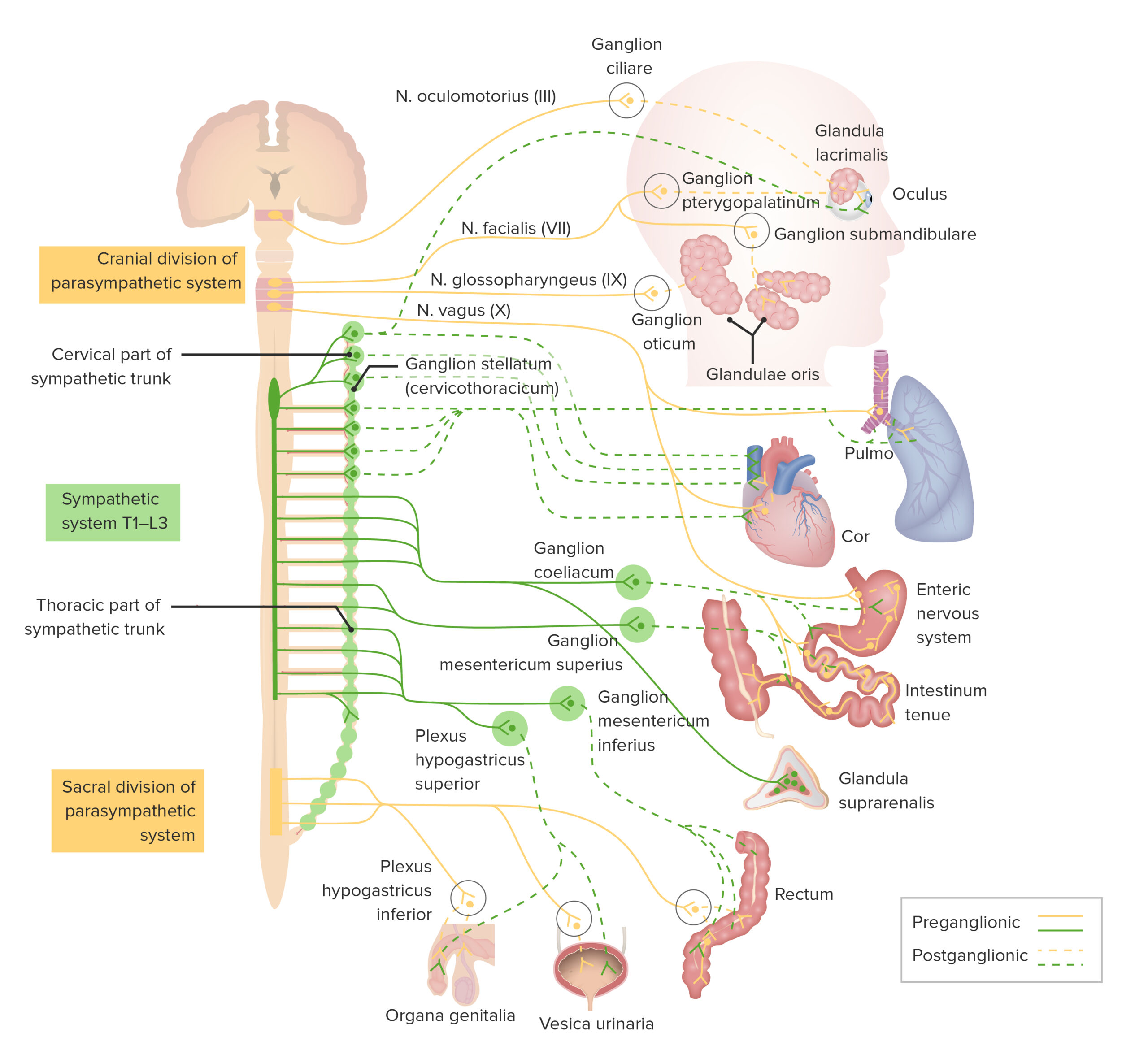
00:01 So if you're a sympathetic neuron, you're leaving the spinal cord from the lateral horn of T1 to L3. 00:08 Ultimately, you're going to pass towards the sympathetic trunk. 00:12 These are sympathetic ganglia that run either side of the vertebral column. 00:17 So sometimes they're known as Paravertebral ganglia. 00:20 Collections of cell bodies in the peripheral nervous system. 00:24 So here we have a sympathetic neuron, the preganglionic neuron passing to the sympathetic trunk. 00:31 It does this by passing to the spinal nerve via the anterior route. 00:36 Once it's in the spinal nerve, it's then going to pass to the sympathetic chain via the white rami communicans. 00:44 Once it's there, it's going to do one of three things. 00:48 The first thing is it forms a synapse, and then passes back to the spinal nerve via the gray rami communicans. 00:58 And it does this so it can then carry on with the spinal nerve and go and supply blood vessels, sweat glands, hair follicles on the surface of the skin. 01:08 So once the preganglionic fiber has passed to the sympathetic chain, via the white rami communicans, do one of three things. 01:17 The first thing is passed back to the spinal nerve via the gray rami communicans to go and supply blood vessels, sweat glands, hair follicles in the skin. 01:27 The second thing it can do is it can actually move up and down the sympathetic chain. 01:33 So although sympathetic nerves only come from T1 to L3, they also need to supply the head and neck and the pelvis. 01:40 And they do this by passing up and down the chain, which runs the entire length of the spinal cord. 01:46 Even though it's only formed by nerves that leave from T1 to L3. 01:52 We'll see that in the next slide. 01:54 So the first thing is to pass the surface of the skin, via the gray gray rami communicans. 01:59 The second thing is to ascend or descend up and down the sympathetic chain. 02:04 And the third thing to do is not actually synapse but move all the way towards what we know as pre-vertebral ganglia. 02:13 And this is why splanchnic nerves. 02:16 And although the preganglionic fiber here is longer than what we've previously described, it is still shorter than the parasympathetic, so we'll see in a moment. 02:26 So if we then look at this slide here, we can see the sympathetic trunk is running either side of the spinal cord. 02:34 The so called paravertebral ganglia. 02:37 These ganglia cervical, thoracic, lumbar, sacral, are formed from sympathetic nerves that lead from T1 to L3. 02:46 But they once they get to the sympathetic trunk, the second option is them to ascend and descend. 02:52 Remember, the first option was to go via the gray rami communicans back to the spinal nerve to go to the surface of the skin. 02:59 The second option now is to ascend and descend up and down the sympathetic trunk. 03:04 And the third one is what we'll see in this next example. 03:08 So here we can now see a series of splanchnic nerves. 03:11 These are carrying preganglionic fibers for from the sympathetic chain. 03:18 These splanchnic nerves pass towards the prevertebral, or preaortic ganglia. 03:24 These sit anterior to the aorta, which sits anterior to the vertebrae, hence prevertebral preaortic. 03:32 But they contain the cell bodies of post-ganglionic fibers. 03:37 So this is where those preganglionic fibers coming from the sympathetic chain via the splanchnic nerves synapse with the postganglionic fibers. 03:47 So let's have a look. 03:49 What we have is the celiac ganglia. 03:52 Now, if you remember, the celiac trunk supplies the foregut. 03:57 The superior mesenteric artery supplies the midgut. 04:01 The renal arteries supply the kidneys and adrenal glands. 04:05 And the inferior mesenteric artery supplies the hindgut. 04:10 We also have branches of the internal iliac supplying the pelvic organs. 04:15 So, why am I telling you this? Running around all of the arteries that emerged from the aorta are periarterial plexi. 04:25 Now these are connections have autonomic nerves that pass to the organs via the blood vessels. 04:31 They piggyback on them. 04:33 So what we have is preganglionic fibers running a splanchnic nerves from the sympathetic chain to their corresponding blood vessel that will take them to their target organ. 04:46 So let's look at the greater splanchnic nerve at the top. 04:50 So the greater splanchnic nerve is going to contain preganglionic fibers destined for the foregut. 04:57 The foregut is supplied by branches of the celiac trunk. 05:01 So the greater splanchnic nerve is taking preganglionic fibers to synapses the celiac ganglion. 05:09 This is then going to give rise to postganglionic fibers that will follow the celiac trunks branches to the foregut. 05:16 The similar happens for the lesser splanchnic nerve to the superior mesenteric ganglion. 05:22 The least splanchnic nerve associated with the aorticorenal ganglion. 05:26 Lumbar splanchnic nerves associated with the inferior mesenteric ganglion and then sacral splanchnic nerves associated with the inferior hypogastric plexus. 05:38 We'll come back to that in a moment. But the principle is this. 05:43 The blood vessels that supply that part of the gut tube foregut, midgut, hindgut are going to receive preganglionic fibers via the splanchnic nerves - greater, lesser, least, lumber. 05:56 They're containing the preganglionic fibers that will sign ups with postganglionic fibers. 06:02 Those postganglionic fibers run to the respective part of the gut tube or the kidneys via periarterial plexi alongside the respective arteries. 06:14 Now, you will see some contradiction in various textbooks around the greater, lesser, least, lumbar splanchnic nerves contribution to each of these. 06:23 Personally, I wouldn't get too worried about that Refer to the specifics in your course if you need to. 06:29 But appreciate, that splanchnic nerves coming away from the synthetic chain will form these ganglia. 06:37 What we can see in the diagram and what is important is connecting the celiac ganglion to the superior mesenteric ganglion, and then the superior mesenteric ganglion, to the inferior mesenteric ganglion, are a series of interconnecting nerves. 06:53 So, there is some connectivity between the celiac ganglion and the superior mesenteric ganglion, and then the superior mesenteric ganglion to the inferior mesenteric ganglion. 07:03 And this is important, because there's a contribution of the sympathetic nerves from these splanchnic nerves down into the pelvis by what's known as the superior hypogastric plexus. 07:17 Now, the superior hypogastric plexus is positioned anterior to the bifurcation of the aorta. 07:24 So, where the aorta bifurcates into the common iliac anterior to it we have the superior hypogastric plexus, which is a continuation down from the inferior mesenteric ganglion. 07:37 Each of the common iliac is then going to be associated, common iliac arteries is going to be associated with a hypogastric nerve. 07:47 So, the superior hypogastric plexus splits into a left and right hypogastric nerve. 07:54 Those left and right hypogastric nerves will then merge with the sacral splanchnic nerves to form the inferior hypogastric plexus. 08:03 Then the hypogastric plexus inferiorly will give rise to periarterial branches alongside the branches of the internal iliac that will go and supply the pelvic organs. 08:16 It is quite complicated, so you may need to listen to that again. 08:20 But the best way to understand this is to try and draw some of these pathways out yourself. 08:24 So, watch the video, listen to the description and try and draw these pathways out for the sympathetics.
About the Lecture
The lecture Sympathetic Nervous System (SNS): Organization by James Pickering, PhD is from the course Lymphatics and Nerves of Abdominopelvic Region.
Included Quiz Questions
What are the destinations for sympathetic preganglionic nerve fibers? Select all that apply.
- Gray ramus communicans
- Sympathetic trunk
- Prevertebral ganglion
- Parasympathetic ganglion
- Black ramus communicans
What is the destination of the splanchnic nerves?
- Prevertebral ganglia
- Sympathetic trunk
- Paravertebral ganglia
- Parasympathetic ganglia
- Pelvic ganglia
Which ganglion is associated with the lesser splanchnic nerves?
- Superior mesenteric ganglion
- Celiac ganglion
- Aorticorenal ganglion
- Inferior mesenteric ganglion
- Obturator ganglion
Which ganglion is associated with the sacral splanchnic nerves?
- Inferior hypogastric plexus
- Inferior mesenteric ganglion
- Aorticorenal ganglion
- Superior mesenteric ganglion
- Celiac ganglion
Author of lecture Sympathetic Nervous System (SNS): Organization

James Pickering, PhD
Customer reviews
5,0 of 5 stars
| 5 Stars |
|
5 |
| 4 Stars |
|
0 |
| 3 Stars |
|
0 |
| 2 Stars |
|
0 |
| 1 Star |
|
0 |
