Playlist
Show Playlist
Hide Playlist
Structures of the Orbit
-
Slides Anatomy Structures of the Orbit.pdf
-
Download Lecture Overview
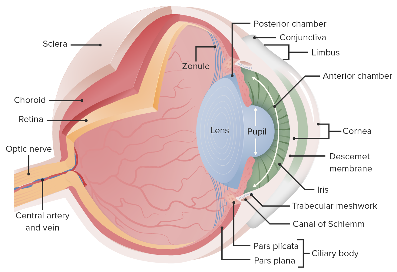
00:01 Let's take a look at some of the other structures found in the orbit. 00:06 Starting with this small little connective tissue sling called the trochlea. 00:11 This trochlea is through which the superior oblique is going to pass before it attaches to the eyeball. 00:19 Here we see the optic nerve carrying visual information from the eyeball and superior and lateral to the eyeball, we have the lacrimal gland or the gland of tear production. 00:31 And again, we have that branch of the ophthalmic nerve the lacrimal nerve in this area. 00:38 While the lacrimal nerve is providing sensory innervation, the parasympathetic innervation is actually carried out by branches going back to the facial nerve. 00:47 So cranial nerve VII is responsible for the parasympathetic innervation of this gland. 00:57 Here we see the superior rectus upon which sits the levator palpebrae superioris, the muscle that acts on the upper eyelid. 01:05 And sitting on top of that, we have that other branch of the ophthalmic nerve, the frontal nerve. 01:14 Here is that frontal nerve from a lateral view. 01:17 Again, here is the lacrimal gland and the levator palpebrae superioris. 01:23 Since we're at a lateral view, we can see the lateral rectus pretty well. 01:28 And we can also see a bit of the inferior rectus and the inferior oblique. 01:36 From an anterior view, we can see the supraorbital nerve exiting out as well as the supratrochlear nerve just a bit medial to it. 01:46 And this anterior view, we can really see the location of the lacrimal gland as being superior and lateral in the orbit. 01:56 And then in the opposite corner, more inferior and medial, we see that nasolacrimal duct which is going to drain the tears. 02:05 They're going to drain the tears via these small canals, hence the term canaliculi called lacrimal canaliculi. 02:14 And again, below the orbit at the infraorbital foramen, we have the infraorbital nerve, which is part of the maxillary division of trigeminal. 02:25 When we look at the eyeball directly, the white part of the eye which is a tough connective tissue layer is called the sclera. 02:33 The circular ring around the center of the eye is the iris. 02:39 And the space at the center of the iris is the pupil.
About the Lecture
The lecture Structures of the Orbit by Darren Salmi, MD, MS is from the course Special Senses.
Included Quiz Questions
What muscle passes through the trochlea?
- Superior oblique
- Inferior oblique
- Superior rectus
- Middle rectus
- Inferior rectus
Which nerve provides parasympathetic innervation to the lacrimal gland?
- CN VII
- CN V
- CN VI
- CN X
- CN IX
Customer reviews
5,0 of 5 stars
| 5 Stars |
|
5 |
| 4 Stars |
|
0 |
| 3 Stars |
|
0 |
| 2 Stars |
|
0 |
| 1 Star |
|
0 |


