Playlist
Show Playlist
Hide Playlist
Structure and Blood Supply of the Kidneys
-
Slides Structure and Blood Supply of the Kidneys.pdf
-
Download Lecture Overview
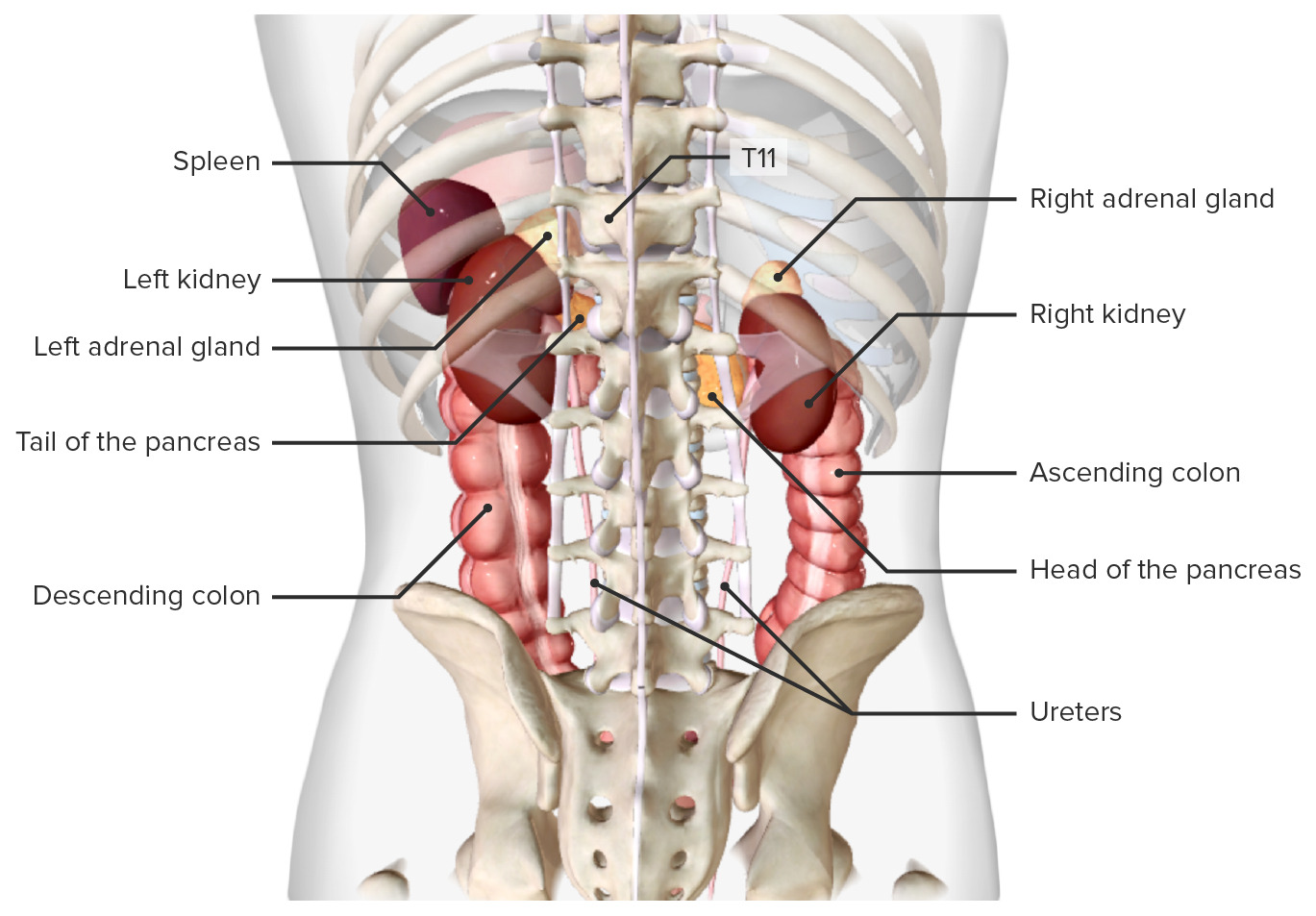
00:01 So now let's specifically look at the kidneys themselves. 00:05 So here we can see the kidneys, and they can be divided up into a large number of segments that helps us locate various different aspects. 00:13 So here we have the superior segment of the kidney. 00:16 We have an anterior superior segment that's projected closer to the anterolateral abdominal wall. 00:22 We also have an anterior inferior segment, and then we have the inferior segment. 00:27 So four different segments there for the kidney, which are important if we think about the blood supply, which we'll come to in a moment. 00:34 We also on the posterior aspect, where you saw an anterior superior and an anterior inferior. 00:41 We have a posterior segment. 00:42 And that's where this kidney has been separated in half down the coronal plane, and we'd have both segments anterior and posterior in that division. 00:53 Now let's look at the right kidney specifically. 00:55 Here we have a smooth anterior surface, which is covered by an easily removable fibrous renal capsule. 01:03 And part of that removal we'll also be clearing that perinephric fat. 01:07 If we were to do a coronal section through the kidney, you will see that on the screen here on the left side of the screen, we have the lateral curved edge, and on the medial surface, we see it's indented. 01:20 At the top of the kidney, we have a superior poll, which obviously means we'll also have an inferior poll projecting at the bottom. 01:27 We have a large convex lateral surface, and we have this concave medial surface indented with the renal sinus. 01:36 Here we can see the renal hilum, which is where blood vessels pass into and leave the kidney. 01:41 And we also have the ureter occupying this space as well. 01:45 The renal sinus is just a space and it's often filled with that perinephric fat. 01:50 And it's really that place where those blood vessels and the ureter as part of the renal pelvis we'll come to in a moment or two occupy this space and then break up into smaller branches. 02:00 We'll look at those branches in a moment or two. 02:03 So here we can see the renal capsule running all the way around the outer layer of the kidney. 02:10 Just deep to that renal capsule, a protective fibrous layer, we have the cortex. 02:15 And that runs around the periphery of the kidney. 02:18 We then have an inner medulla and these are full of these kidney pyramids. 02:23 They are associated with the processing of urine as the blood flows through the various aspects of the kidney. 02:31 The human is then collected and passed out by way of the ureter. 02:36 Here we see one of those renal pyramids with its apex projecting to the renal sinus. 02:41 We have a base, a nice broad flat base of the renal pyramid and we have an apex. 02:46 And here there was an opening called a renal papilla. 02:50 And that is where urine passes into what are known as minor calyx. 02:54 Here we can see one minor calyx. 02:57 So each renal pyramid will give rise to a renal papilla where urine passes and enters into its minor calyx. 03:05 Here we can see the cortex that surrounding the kidney again, just deep to that fibrous layer. 03:11 And parts of that cortex actually passes between the renal pyramids, and these are known as renal columns. 03:18 If we have a slightly closer look, we can see that the renal pyramid renal papilla gives rise to those minor minor calyces that begin collecting the urine. 03:28 One two or three of these minor calyces can actually drain into a major calyx and the kidney will have about three or four of these major calyces with each of those major calyces receiving two, three, or four minor calyces. 03:45 So the renal pyramid helps to produce the urine. 03:48 It passes it through the renal papilla into the minor calyx or collecting area, and that minor calyx will run into a major calyx and a number of minor calyces will form a major calyx. 04:02 Once those major calyces have united, they form what's known as a renal pelvis. 04:07 And that's the largest of these collecting areas. 04:10 The renal pelvis then funneled down into the ureter, which will merge with the bladder, and we'll see that later on. 04:17 So here we can see the two kidneys in situ. 04:20 We can see them in position associated lateral to the aorta. 04:24 We can see the ureter leaving through the renal sinus through the renal hilum and also passing into the kidney, we can see the two renal arteries. 04:34 So here we can see the left renal artery passing to the left kidney. 04:38 And here we can see the right renal artery passing towards the right kidney. 04:43 Sitting next to the aorta and sitting anterior to these rinus, to these arterial structures, we find the inferior vena cava. 04:52 And coming away from each of the kidneys, we have the right renal vein here, and the left renal vein. 04:58 And these are sitting anterior to the arteries. 05:02 Let's have a close up look of the vascular supply to the kidney. 05:06 And this can actually be quite complicated. 05:09 So let's maybe take it slower and go through these individually. 05:12 So here we see our renal artery. 05:15 Now the renal artery is going to give a posterior branch which remember goes to that posterior segment of the kidney. 05:23 So there we've got a posterior branch. 05:24 And if we've got a posterior branch, we're going to have an anterior branch. 05:28 Here's the anterior branch, bifurcating. 05:31 It gives an apical segmental artery that heads towards the apex of those renal pyramids. 05:37 Here we can see part of the segmental artery passing away to the respective areas. 05:43 He is the anterior, superior, and inferior segmental arteries. 05:48 They are the arteries supplying those specific segments of the kidney. 05:54 We then see the inferior segment artery heading down to the bottom aspect, the inferior aspect of the kidney. 06:02 A closer look looks at how these blood vessels work around the renal pyramids. 06:06 So here we have a segmental artery passing between the renal pyramids. 06:10 It gives these interlobar arteries that run between the renal pyramids. 06:15 They're running through the renal columns, that extension of cortex that pass between the renal pyramids. 06:22 Once they get there, they give these arcuate arteries that run around the base of the renal pyramids. 06:28 And here again, we can see some smaller interlobar arteries running through the segments in the parenchyma the substance of the kidney to supply the various pieces of tissue. 06:39 These blood vessels are really important in helping to take blood to the kidney and allowing it to be filtered and allow urine and the water balance to be maintained as the primary function of the kidney. 06:52 The blood supply to these region then allows urine to pass through the papilla, minor calyx, major calyces, into the renal pelvis and then the ureter. 07:02 So here we see the ureter is sitting most posteriorly within this space, then we have the renal artery, and then we have the renal vein most anteriorly. 07:11 So if you're looking at anterior kidney, the first thing you'll see is the renal vein. 07:16 Then you'll have a look at the renal artery and then you'll see the ureter most posteriorly.
About the Lecture
The lecture Structure and Blood Supply of the Kidneys by James Pickering, PhD is from the course Anatomy of the Urinary System and Suprarenal Glands.
Included Quiz Questions
Where are nephrons located in the kidney?
- Cortex and pyramids
- Major calyx
- Minor calyx
- Renal cortex
- Renal column
Which structure is located outside the renal sinus?
- Ureters
- Renal arteries and veins
- Fat
- Major calyces
- Nerves
Customer reviews
5,0 of 5 stars
| 5 Stars |
|
5 |
| 4 Stars |
|
0 |
| 3 Stars |
|
0 |
| 2 Stars |
|
0 |
| 1 Star |
|
0 |


