Playlist
Show Playlist
Hide Playlist
Scapula

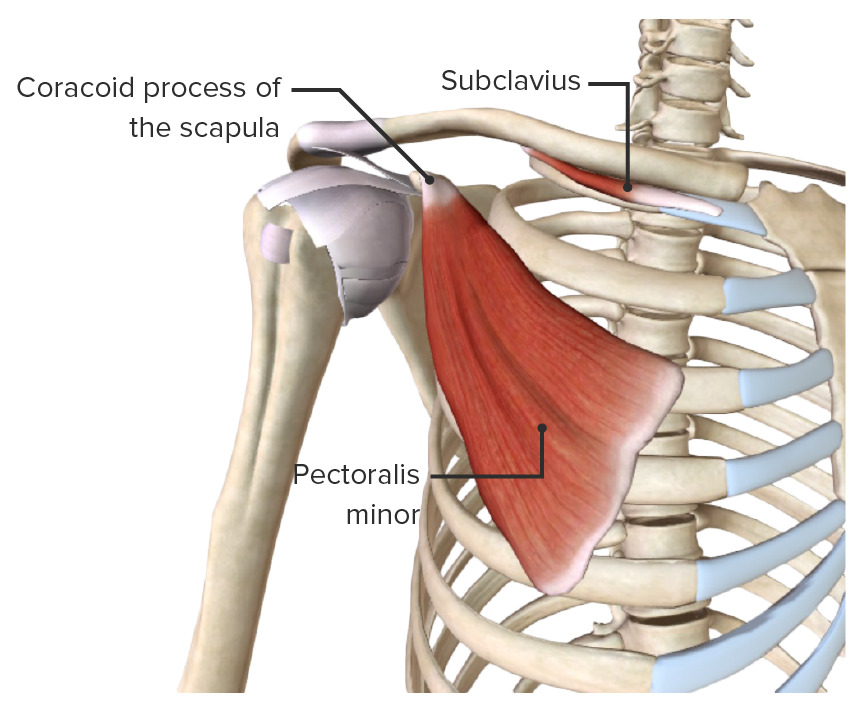
00:01 So, now, let's turn our attention to the scapula. 00:04 So, let's just have a look at where the scapula is located. 00:08 It's on the posterior aspect of the thoracic cage and we can see it here highlighted in green. 00:14 The only bony attachment it has with the axial skeleton is via the clavicle which attaches to the sternum. 00:21 So, remember, the only way the axial skeleton is in continuation with the appendicular skeleton is by that sternoclavicular junction, that sternoclavicular joint and here, we can see them projecting all the way over to the scapula. 00:36 The scapula you can see there is associated with the second and the seventh rib. 00:40 To give you the extent of its superior and inferior boundaries. 00:44 So, here, we can have a look at the scapula. This is the costal surface. 00:48 So, here, we're looking at a right scapula. 00:51 And we're looking at the surface that is tightly adhered or adjacent to the costal surface of the ribs. 00:58 We can see if we then turn it around, we're looking at the posterior surface and we have what sometimes is called the dorsal surface. 01:06 So, the costal surface or the anterior surface. And here, we're looking at it on its posterior aspect. 01:13 Here, we can look back at the anterior aspect. We see there is a superior border. 01:17 There is an angled lateral border and we have a medial border that will be running next to the vertebral column. 01:24 We can also see most superiorly where the superior border and the medial border unite. 01:30 There's a superior angle. And here, where the medial and lateral borders unite, we have an inferior angle. 01:38 Where there is the most lateral extremity, we call this bit the lateral angle. 01:44 Now, the scapula is going to be really popular with lots of muscles. 01:50 There's lots of muscle attachments that are situated here and that actually helps to adhere the scapula tight to the costal surface of the ribs. 01:59 So, it helps to pull it against the thoracic cage. Here, we can now see the posterior view with the scapula being rotated and we have this really prominent ridge that runs across the posterior surface. 02:11 We have the spine and then, it becomes dilated as the acromion. 02:15 And remember, the acromion was in continuation with the clavicle and the acromioclavicular joint. 02:22 So, here, we can see the spine of the scapula, the acromion. 02:26 We also have another bony prominence that kind of twists and extends out from the scapula. 02:32 This is known as the coracoid process. 02:35 And these bony regions are important for enhancing the opportunities for muscle to attach to the skeleton. 02:42 So, let's have a look at this costal surface in a bit more detail. 02:46 Again, this is a right scapula and we're looking at it as if we're looking through the thoracic cage. 02:52 Here, we have a slight shallow indentation which is a subscapular fossa. 02:57 It's home to an important muscle, subscapularis. 03:00 And then, if we rotate the scapula around onto its dorsal surface, again, we can see the spine of the scapular and above the spine, we have two fossae. 03:10 We have the supraspinous fossa above and we have the infraspinatus fossa below. 03:16 So, the spine of the scapular separates that region into two fossae, supraspinous and infraspinatus. 03:23 Connecting the two fossae, we have the spinoglenoid notch, an important part where blood vessels can pass through this region and it also helps to connect those two fossae. 03:34 You'll see along the superior border of the scapula, we have this little indentation. 03:39 That's known as the suprascapular notch. This has a little roof on it which is known as the superior transverse ligament or the suprascapular ligament And this creates the suprascapular foramen or the suprascapular canal. 03:54 This is an important space as it allows structures to pass through it It predominantly has the superscapular nerve running through it which goes on the supply muscles around the scapula And in some cases it will also have the suprascapular atery running within the canal Although most of the time this artery is located superficial to it. 04:14 If this canal was to ossify during the ossification process of that superior transverse ligament Then it could compress the suprascapular nerve and lead to the associated paralysis. 04:27 Let's now look at the medial border of the scapula. 04:30 And again, this is going to be home to lots of muscle attachments that will attach to this region. 04:36 So, the medial border here most superiorly, we have an attachment for levator scapulae. 04:41 Here, we have an attachment site for rhomboid minor. 04:44 And then, inferior to rhomboid minor with our rhomboid major. 04:48 And these are going to extend either to the neck, levator scapulae or to the vertebral column as the rhomboid muscles. 04:55 We also have a series of muscles coming away from this lateral border. 04:59 Most inferiorly, we have teres major. And then, we have teres minor more superiorly. 05:05 Coming inferiorly down from this region, we have the long head of triceps. 05:10 We'll come back to this region when we talk about the triceps in more detail. 05:14 But these are important muscles, some of which go on to form the rotator cuff. 05:19 If we look at the superior angle and the inferior angle, we can now see these on this posterior view of the scapula and again, we can see the lateral angle, important locations for those muscles which were rising from the scapula. 05:35 Now, we're going to have a look at the scapula as if it's from the lateral aspect. 05:41 So, we've taken away the humerus that would sit on this circular region and we're looking at it from its lateral aspect. 05:47 So, if someone's standing by your side and looking at your scapular side on. 05:51 The most prominent feature we can see here is that circle structure I eluded to and this is the glenoid fossa. 05:58 The glenoid fossa we can see is going to be home to the head of the humerus. 06:03 We'll come to that in a moment. Just medial to the glenoid fossa, we can see the scapular neck and you can see that highlighted here in green and then, an alternative view of the coracoid process and the supraglenoid tubercle. 06:17 The supraglenoid tubercle is important as it offers another bony attachment site and we can see the supraglenoid tubercle is also going to be paired with the infraglenoid tubercle. 06:28 These two tubercles sitting above and below the glenoid fossa. 06:32 Now, we're going to have a look at the superior view of the scapula. 06:37 We can see that on the right-hand side. 06:39 So, towards the right of the screen, we have the medial border and then, towards the left-hand side, we have the lateral aspect where we can see the acromion, the coracoid process and parts of the glenoid fossa. 06:50 So, here, we can see that lateral border that we've spoken about before. 06:53 And here, we can see the anterior border going on that posterior aspect of the scapula. 06:59 This is the spine of the scapula that we're familiar with. 07:03 If you look at this aspect from the top down, you can now see that the side of the scapula has this superior surface, an important location for supraspinatus muscle. 07:13 And here on the underside, you can see the inferior surface which is an important landmark for infraspinatus muscle. 07:20 Again, offering important bony attachment sites for various muscles. 07:26 Here, we can now see how the supraspinatus muscle occupies this space. 07:30 It will pass out laterally to associate itself with the head of the humerus. 07:36 As we have a supraspinatus muscle, we're going to have an infraspinatus muscle and that's occupying this space inferior to the spine of the scapula. 07:45 Surrounding all of this area and most superficial of all of the muscles is deltoid. 07:50 This is an important muscle that runs all the way around posteriorly on the spine of the scapula. 07:56 It also goes around the acromion and helps to surround a large proportion of the shoulder joint. 08:03 We can also have running medially from the spine of the scapula, another large and very fanned out muscle, trapezius which we can see here. 08:12 Don't worry too much about these muscles at the moment. 08:15 We'll cover them in a lot more detail as we progress through this course. 08:21 So, let's just finish up by looking at some various other parts of the scapula. 08:25 Here, we have the acromion, we have the acromial angle. 08:28 And here, we can now see how the clavicle, introduced into the acromion, forms the acromioclavicular joint. 08:35 Here, we have the coracoid process, again, another bony outgrowth that helps to offer muscle attachments and we'll come to that later on.
About the Lecture
The lecture Scapula by James Pickering, PhD is from the course Osteology and Surface Anatomy of the Upper Limbs.
Included Quiz Questions
Which of the following statements regarding scapula is correct? Select all that apply.
- The anteromedial border of the scapula lies directly posterior to ribs 2–7.
- The glenoid cavity articulates with the clavicle.
- The glenohumeral joint is an articulation between the head of the humerus and a specialized region of the scapula.
- The coracoid process is located on the lateral aspect of the superior border of the scapula.
At which thoracic rib level is the scapula located?
- 2–7
- 1–7
- 3–7
- 4–7
- 1–5
Which border connects the superior angle of the scapula with the inferior angle?
- Medial
- Lateral
- Superior
- Posterior
- Inferior
Which dislocation of the humerus does the acromion prevent?
- Superior
- Anterior
- Posterior
- Inferior
- Lateral
The fossa on the anterior surface of the scapula provides attachment to which muscle(s)?
- Subscapularis
- Supraspinatus
- Infraspinatus
- External intercostal muscles
- Brachioradialis
Author of lecture Scapula

James Pickering, PhD
Customer reviews
5,0 of 5 stars
| 5 Stars |
|
5 |
| 4 Stars |
|
0 |
| 3 Stars |
|
0 |
| 2 Stars |
|
0 |
| 1 Star |
|
0 |
