Playlist
Show Playlist
Hide Playlist
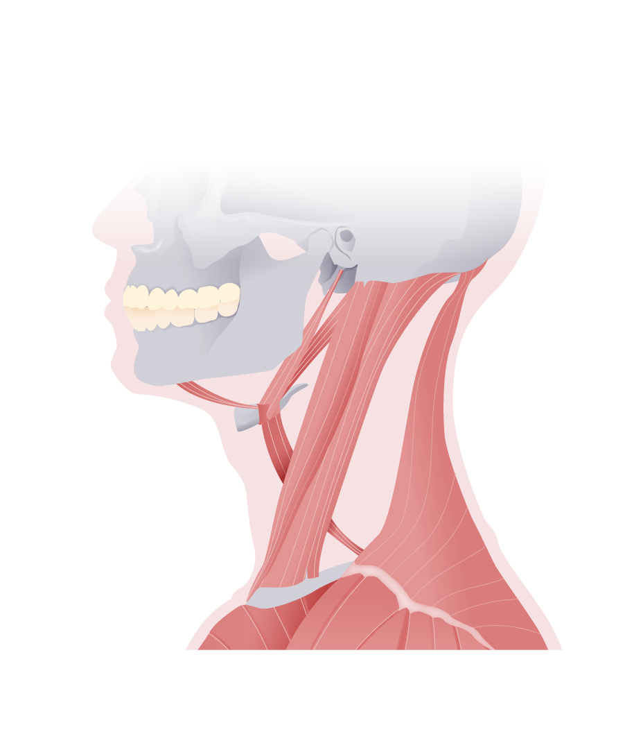
Posterior Triangle of the Neck

-
Slides Anatomy Posterior Triangle of the Neck.pdf
-
Download Lecture Overview
00:01 Now that we looked at the anterior triangle of the neck, we're going to look on the other side of the sternocleidomastoid to find the posterior triangle. 00:11 So the boundaries of the posterior triangle are the posterior edge of the sternocleidomastoid, the trapezius muscle, and the middle third or so of the clavicle. 00:24 The posterior triangle has what we consider a floor, which are the muscles deep to this area, which are the splenius, capitis, levator scapulae, and the three scalene muscles posterior, middle, and anterior. 00:39 We can divide the posterior triangle a little bit more, but really only into two smaller ones. We can divide it on either side of the inferior belly of the omohyoid, whereas inferior to that we will have the omoclavicular or subclavian triangle, and superior to it would be the occipital triangle. 00:59 Let's look at some of the contents of the posterior triangle, starting with the external jugular vein. 01:06 External refers to its location, external to the sternocleidomastoid. 01:11 And here we see that external jugular vein coming down into the subclavian vein where it drains. We also see posterior to that the subclavian artery. 01:23 And if we fade out the sternocleidomastoid, we see the accessory nerve, which is what innervates the sternocleidomastoid and trapezius muscles. 01:32 We also see this very important plexus of nerves called the cervical plexus, as well as a collection of nerves that are going to supply the upper limb called the brachial plexus. So let's look again at that external jugular venous drainage. 01:51 In terms of venous drainage of the face, the retromandibular vein does the majority of the work and it has an anterior and posterior division. 01:59 In this case, we're going to look at that posterior branch that merges with the posterior auricular vein coming from behind the ear to actually form the external jugular vein, which will in turn drain into the subclavian vein. 02:15 Now let's take a look at the subclavian artery. 02:20 Here we see the subclavian artery arising from the brachiocephalic trunk on the right side. On the left, the subclavian artery arises directly from the aortic arch owing to the asymmetry of this area. 02:35 After the subclavian artery passes the outer border of the first rib, it's going to change name to the axillary artery. 02:42 And eventually, that will even change its name to the brachial artery as it enters the upper limb. 02:48 If we zoom in a little bit more. 02:50 Here we see this muscle called the anterior scalene muscle. 02:55 And this is the muscle behind which the subclavian artery is going to pass anterior to the scalene muscles where we would find the subclavian vein. 03:06 The subclavian artery here can be divided into the prescalene part or the portion proximal to the anterior scalene. 03:13 The retroscalene part, the part that's sitting just behind it. 03:17 And then the short postscalene part just before it turns into the axillary artery. 03:26 In terms of the prescalene part, we have the vertebral artery and that's a very important artery in this area because this is the artery that's going through the transverse foramina of the cervical vertebra on its way up into the brain to form a very important anastomosis in the circle of Willis. 03:43 At about this same area but traveling inferiorly would be the internal thoracic artery, very important artery for the thorax. 03:52 We also have the thyrocervical trunk, which is a short trunk that branches into the suprascapular artery, the transverse cervical artery, and the inferior thyroid artery. 04:05 The superior will be a branch of the external carotid. 04:10 In terms of the retroscalene part, we have the costocervical trunk, which is going to give rise to the deep cervical artery, the supreme intercostal artery, which is just the uppermost intercostal artery, and the dorsal scapular artery. 04:27 Now let's take a look at the nerves in this area. 04:30 They come from a plexus called the cervical plexus. 04:33 Here we see the cervical spinal roots C1, C2, C3, and C4. 04:39 Several cutaneous nerves come off of this area, including the lesser occipital, great auricular, transverse cervical, and the supraclavicular nerve. 04:51 We also see C1, C2, and C3 forming this loop called the ansa cervicalis. 04:58 And this is going to provide motor supply to the infrahyoid muscles, with the exception of thyrohyoid, which is only receiving motor innervation from C1. 05:09 If we throw in C5 as well, we see the origins of the phrenic nerve, which is composed of roots from C3, C4, and C5 before it goes all the way down to supply the diaphragm. 05:23 So here are those cutaneous branches. 05:26 We see the lesser occipital going in the area of the occipital bone, the great auricular in the general area of the ear, the transverse cervical and supraclavicular nerves. 05:37 And as you can see, they're all mostly in the posterior area of the head and neck. 05:42 And that's because the anterior portion of the head gets its cutaneous innervation from the trigeminal nerve.
About the Lecture
The lecture Posterior Triangle of the Neck by Darren Salmi, MD, MS is from the course Neck Anatomy.
Included Quiz Questions
What is the posterior border of the posterior triangle?
- Trapezius
- Sternocleidomastoid
- Anterior third of the clavicle
- Middle third of the clavicle
- Posterior third of the clavicle
Which structure divides the posterior triangle into the subclavian and occipital triangles?
- Inferior belly of the omohyoid
- Trapezius
- Sternocleidomastoid
- Anterior third of the clavicle
- Posterior third of the clavicle
Which structure is NOT part of the posterior triangle?
- External jugular artery
- External jugular vein
- Subclavian artery
- Subclavian vein
- Accessory nerve
Author of lecture Posterior Triangle of the Neck

Darren Salmi, MD, MS
Customer reviews
5,0 of 5 stars
| 5 Stars |
|
1 |
| 4 Stars |
|
0 |
| 3 Stars |
|
0 |
| 2 Stars |
|
0 |
| 1 Star |
|
0 |
1 customer review without text
1 user review without text
