Playlist
Show Playlist
Hide Playlist
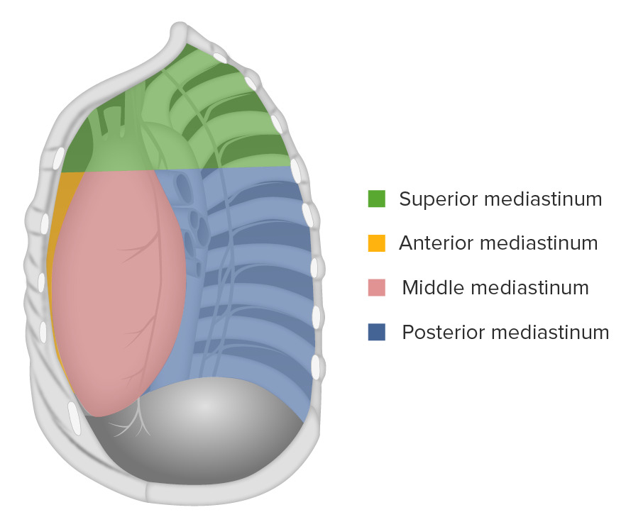
Posterior Mediastinum

-
Slides Anatomy Posterior Mediastinum.pdf
-
Download Lecture Overview
00:01 Although we've already seen the mediastinum. 00:03 We're going to revisit the posterior mediastinum. 00:06 Starting with some superficial stuff, then some deep stuff. 00:09 Now that we've had a greater understanding of the rest of the mediastinum. 00:15 So let's go back and look at the vagus nerves. 00:19 So the vagus nerves, as we know are descending from the neck area down into the thorax, and eventually into the abdomen. 00:25 And here we see the right vagus nerve, and the left vagus nerve. 00:30 And we also know that there are what are called recurrent branches. 00:34 Recurrent laryngeal branches that we're going to deal with. 00:37 But before we see those, we know that distally they're going to form a plexus over the esophagus called the esophageal plexus. 00:46 But that recurrent laryngeal that we saw is wrapping under the aortic arch on the left. 00:52 But the aortic arch is an asymmetric structure. 00:54 So let's look at this in a little bit greater detail. 00:58 On the right side, we see the right vagus nerve coming down to the level of the right subclavian artery. 01:05 And then if we blink that out, we can actually see the right recurrent laryngeal nerve here wrapping underneath the right subclavian artery to head back towards the trachea in the larynx. 01:17 But on the left side is quite a bit different. 01:19 We have the left vagus nerve coming down a lot further before it gives off the left recurrent laryngeal nerve. 01:28 And that's because of embryology essentially. 01:32 The recurrent laryngeal nerve started out very symmetric early in development, but because of regression of some right sided structures, and ended up higher up underneath the subclavian. 01:42 But on the left side, we had that thing called the ductus which becomes ligamentum arteriosum. 01:48 And it sort of traps the left recurrent laryngeal nerve underneath the aortic arch. 01:55 Now, let's look at some deep structures of the posterior mediastinum. 02:01 If we look deeply into the posterior mediastinum, we see a lot of important veins. 02:06 We already know that the IVC is bringing deoxygenated blood from below the heart. 02:12 And the SVC is bringing deoxygenated blood from above the heart. 02:17 What about the area in between? That's something we have to deal with. 02:21 Importantly, we have intercostal veins in between. 02:24 And what they're going to do is drain into the Azygous veins. 02:28 And on the right side, they can drain directly into the azygous because it's a right sided structure. 02:34 But on the left, they have to do something a little different. 02:37 Inferiorly, they're going to drain into the Hemiazygous vein. 02:41 And then superiorly into the accessory hemiazygous vein, and those will cross over the midline, over to the right side to reach the azygous vein. 02:51 If we look at a lateral view, we can see the arrangement of the SVC receiving this azygous vein from posterior. 03:02 And there's also an exception to this rule, generally speaking, where the first also called supreme or highest intercostal artery takes a shortcut and instead drains directly into the brachiocephalic vein instead of the azygous vein Another important deep structure of the posterior mediastinum are the paired sympathetic trunks going along either side of the vertebral column, in between which will find a lymphatic structure called the thoracic duct. 03:33 This thoracic duct is going to go all the way through the thorax up towards some venous structures. 03:41 Notably, it's going to join at the junction of the left internal jugular vein and the left subclavian vein, so that all that lymphatic fluid it's draining can enter the venous system. 03:53 So again, we have the thoracic duct draining most of the lymphatic fluid, especially all of this stuff coming from the abdomen. 04:01 But we have a smaller duct on the right simply called the right lymphatic duct. 04:06 And it's draining into the same location but on the right side. 04:11 So what this does is essentially drain the right upper part of the body, including the right upper limb, right thorax, and right head and neck. 04:21 Whereas, the thoracic duct handles essentially everything else.
About the Lecture
The lecture Posterior Mediastinum by Darren Salmi, MD, MS is from the course Thorax Anatomy.
Included Quiz Questions
The left recurrent laryngeal nerve curves inferiorly around which structure?
- Aortic arch
- Pulmonary trunk
- Azygos vein
- Hilum
- Subclavian artery
The azygos vein drains into which structure?
- SVC
- IVC
- Right atrium
- Left atrium
- Pulmonary trunk
Author of lecture Posterior Mediastinum

Darren Salmi, MD, MS
Customer reviews
5,0 of 5 stars
| 5 Stars |
|
5 |
| 4 Stars |
|
0 |
| 3 Stars |
|
0 |
| 2 Stars |
|
0 |
| 1 Star |
|
0 |
