Playlist
Show Playlist
Hide Playlist
Heart in Situ
-
Slides Anatomy Heart in Situ.pdf
-
Download Lecture Overview
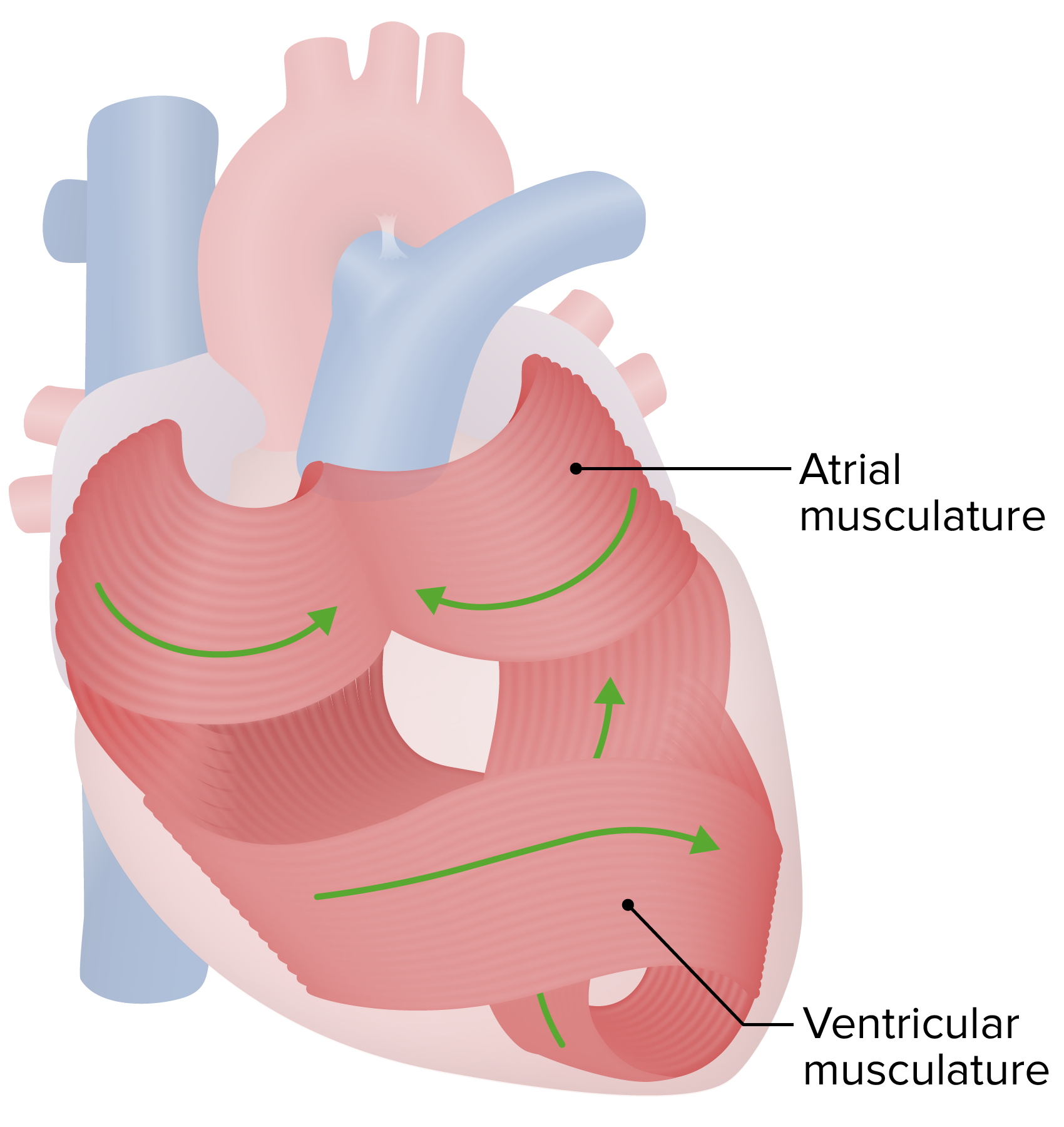
00:01 Alright, now we've reached the heart of the course, quite literally. 00:05 We're finally going to talk about the heart. 00:08 It's an organ that probably needs no introduction, but we're going to give it one anyway. 00:12 We're going to start with some very basic terms to describe the heart and some external features. 00:18 We're actually going to start with some terms very similar to the lung, because the heart has a base, and it has an apex. 00:26 And just like the lung, the base is the broader part, and the apex is the pointer part. 00:31 But you notice it's kind of upside down from what we saw in the lungs. 00:34 Here, the apex is actually pointing down instead of pointing up as they were in the lungs. 00:39 You've probably heard of the chambers of the heart. 00:41 But let's see if we can identify them externally. 00:44 We're going to go in the order that a deoxygenated red blood cell would probably follow as it's going through the circulatory system. 00:53 So, we'll start with the right atrium. 00:55 We see the right atrium here, sitting just posterior to the right ventricle. 01:02 And we sing around to the other side to see the left. 01:05 We have the left atrium, again more posterior to its corresponding left ventricle. 01:12 And we can have some idea of where the heart sits, because we talked about the mediastinum. 01:17 And we know it's going to be in the mediastinum. 01:20 So let's look at some of those relations in a little bit greater detail. 01:23 So we have the right lungs on one side, the left lungs on the other, and the sternum, blocking our view of it anteriorly. 01:34 So in order to actually see the heart, we're going to have to remove the sternum. 01:39 Now we can see the heart better. 01:41 And we can see that it doesn't sit perfectly centered in the middle of the thoracic cavity. 01:45 Instead, it's mostly shifted and pointed off to the left side. 01:50 And that's why there's only two lobes on the left lung, where there are three lobes on the right lung. 01:58 And this little portion of the upper lobe that drapes just over the anterior surface of the heart, is called the lingula. 02:09 It gets a little special name because it is this little tongue of lung that sits over the anterior surface of the heart. 02:16 In fact, that's what lingua means. 02:18 It means little tongue, so it's quite literally the little tongue of the lung. 02:21 And where there's really no room for the lung is this knotch called the cardiac knotch. 02:28 Inferiorly we have the diaphragm, which is fused to the pericardium in this region. 02:34 Now, let's swing around to the right side and see some of these relationships. 02:39 Again, inferiorly, we have the diaphragm through which the IVC is coming to join the right atrium, and then posteriorly, we have the descending aorta, and the esophagus, both passing through the diaphragm as well. 02:54 Then we have the trachea. 02:57 Although, it is bifurcating right at the level of these pulmonary vessels, because that's where they're going to enter the lung together. 03:04 And this relationship of the esophagus to the posterior surface of the heart is pretty important to know, because that's why certain types of heart imaging or echocardiography are best done using an esophageal probe. 03:17 And those esophageal probes can give you a better idea of structures that sit posteriorly such as the atria.
About the Lecture
The lecture Heart in Situ by Darren Salmi, MD, MS is from the course Thorax Anatomy.
Included Quiz Questions
Where does the inferior vena cava drain?
- Right atrium
- Left atrium
- Left ventricle
- Right ventricle
- Lingula
What is the left-sided anatomic parallel of the right middle lobe of the lung?
- Lingula
- Cardiac notch
- Pulmonary ligament
- Hilum
- Thymus
Customer reviews
5,0 of 5 stars
| 5 Stars |
|
5 |
| 4 Stars |
|
0 |
| 3 Stars |
|
0 |
| 2 Stars |
|
0 |
| 1 Star |
|
0 |


