Playlist
Show Playlist
Hide Playlist
External Ear

-
Slides Anatomy External Ear.pdf
-
Download Lecture Overview
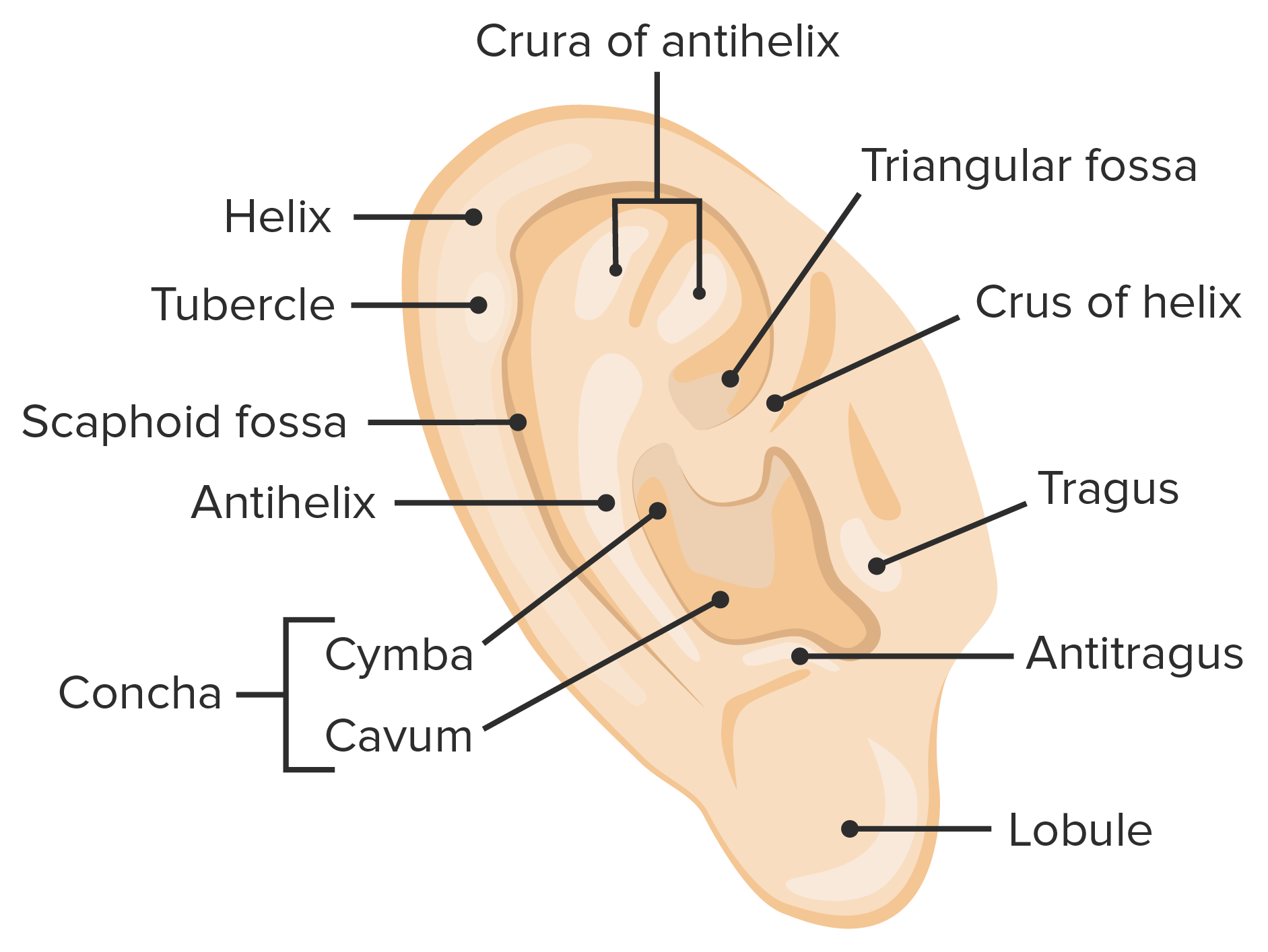
00:01 Now let's talk about the anatomy of the ear. 00:05 The ear is a lot more than the portion you can see. 00:09 We're going to see that the ear has multiple parts, an external, a middle and an inner part. 00:15 Each part getting a little more complex as we go. 00:18 Again, we have an external component, a middle component, and all the way embedded in bone, an internal ear. 00:27 We'll start though with the external component that you're probably most familiar with. 00:32 The external portion that you can see is the auricle or pinna. 00:38 And it will lead to the ear canal and terminate at the eardrum or tympanic membrane. 00:49 We can name some of the parts of the auricle or pinna, such as the interior most flap that we call the tragus, which is separated by a small distance from the antitragus. 01:04 Along the outer edge, we have a ridge called the helix. 01:08 And then a shallow depression separates that from a smaller, more internally located, antihelix. 01:16 Finally, in the center, we have a depression called the concha. 01:20 That's going to lead to our external acoustic meatus the opening for our ear canal. 01:28 Inferiorly, we have a soft portion called the lobule, just an area that's commonly pierced. 01:35 If we take off the skin, we can see the auricular cartilage, which is made up of elastic cartilage, which is just what it sounds like cartilage with the addition of elastin fibers. 01:46 That makes the external ear much more deformable than other types of cartilage that are more rigid. 01:53 We also have extrinsic and intrinsic auricular muscles, whether they're outside or on the ear itself. 02:01 But in humans, they don't really do much as opposed to some other mammals that are able to move their ears and better direct to how they hear things. 02:13 And again, in the center of all this, we have our external auditory canal. 02:21 The arterial supply to the auricle largely comes from the external carotid artery, namely some branches of the superficial temporal artery called the anterior auricular arteries, as well as another branch of the external carotid, the posterior auricular artery. 02:40 In terms of sensory innervation, we have a lot of different nerves contributing. 02:47 We have the lesser occipital, the greater auricular nerve. 02:51 And then we also have some cranial nerve sensation. 02:54 We have a auriculotemporal, off of the mandibular branch of trigeminal. 03:01 And we also have smaller contributions as we get deeper into the auricle or pinna, from other cranial nerves such as cranial nerve X, the vagus nerve, and cranial nerve VII, the facial nerve. 03:15 Here's a cross section showing the pathway of the ear canal. 03:20 So we start with the external auditory canal, which has the cartilaginous part most externally before getting into the bone of the skull through the bony part and will terminate at the tympanic membrane. 03:36 And here is the tympanic membrane, the sort of view you would have through an otoscope into a patient's ear. 03:44 In particular, this would be looking at a patient's right eardrum. 03:49 Our landmarks here would be this little knob called the umbo, which is the end of the handle of a middle ear bone called the malleus. 04:00 This prominent projection here is called the lateral process of the malleus. 04:06 And superior to that, we call that portion of the eardrum, the pars flaccida, and the larger portion more inferiorly the pars tensa. 04:14 And we can see we have these little ridges called the posterior and anterior malleolar folds between the two. 04:23 You'll also notice this little area here called the cone of light, and it's this area where light will bounce back and look pretty bright under an otoscope. 04:33 And it will sit about at the five o'clock position in a right ear or in the left ear about seven o'clock. 04:41 This brings us to how we break up the tympanic membrane when we're describing where a pathology might be located. 04:49 So we break it up into four quadrants. 04:51 We have an anterior superior quadrant, an anterior inferior quadrant, a posterior inferior quadrant and a posterior superior quadrant. 05:03 Now here is an otoscopic view of a patient's tympanic membrane where we can see some of those landmarks. 05:10 Again, we have this inferior portion of that middle ear bone called the umbo. 05:16 And then it's attached to the handle of the malleolus. 05:20 This projection here more superiorly is the lateral process. 05:25 And above that is the parse flaccida. 05:27 Below that, the majority of the membrane is the pars tensa. 05:31 And we can see that this must be a patient's right eardrum based off the location of the cone of light. 05:39 We can also see the structure passing just behind the eardrum. 05:43 And this is actually the chorda tympani or it's a little tiny branch of the facial nerve. 05:49 Hence, its name chorda tympani, because it's passing behind the tympanic membrane.
About the Lecture
The lecture External Ear by Darren Salmi, MD, MS is from the course Special Senses.
Included Quiz Questions
What is the name of the cartilaginous projection anterior to the external opening of the ear?
- Tragus
- Helix
- Antihelix
- Acoustic meatus
- Concha
Which nerve does NOT provide sensation to the external ear?
- Hypoglossal nerve
- Lesser occipital nerve
- Greater auricular nerve
- Mandibular nerve
- Vagus nerve
What marks the end of the external auditory canal?
- Tympanic membrane
- Helix
- Tragus
- Antihelix
- Antitragus
What is the location of the cone of light in the right ear?
- 5 o'clock
- 1 o'clock
- 3 o'clock
- 10 o'clock
- 11 o'clock
Author of lecture External Ear

Darren Salmi, MD, MS
Customer reviews
5,0 of 5 stars
| 5 Stars |
|
5 |
| 4 Stars |
|
0 |
| 3 Stars |
|
0 |
| 2 Stars |
|
0 |
| 1 Star |
|
0 |
