Playlist
Show Playlist
Hide Playlist
Embryologic Development of the Heart and Circulation

-
Slides Anatomy Embryologic Development of the Heart Circulation.pdf
-
Download Lecture Overview
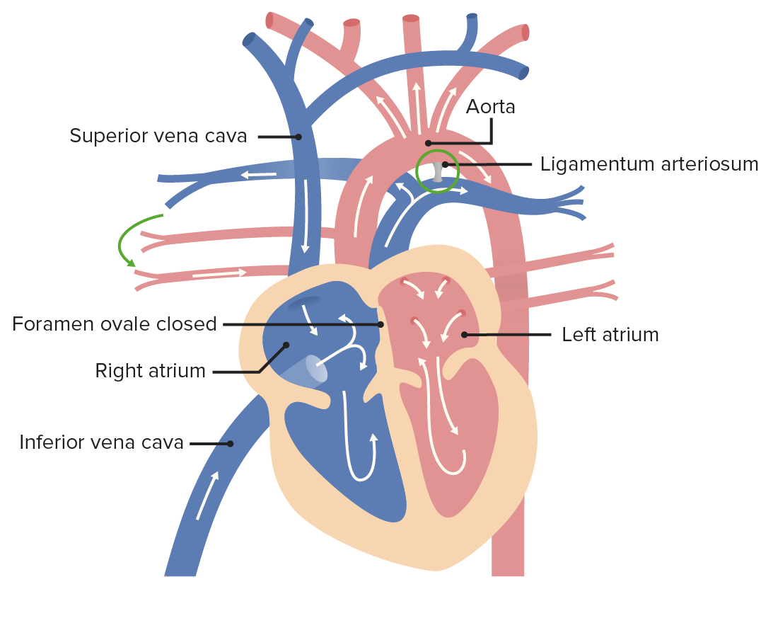
00:01 Now, before we move on to the actual anatomic structures in greater detail, we're going to talk about the big picture of circulation. 00:08 That's going to help us understand the anatomy to come in the following sections a lot better. 00:13 Of course, in order to know that we actually have to talk about how circulation differs before and after birth. 00:20 So we're going to start with fetal circulation. 00:23 Because in fetal circulation, oxygenated blood is actually coming from the placenta, to the body through the umbilical vein. 00:33 And I'll stop right there because you might be wondering, "Why is the vein carrying oxygenated blood? I thought arteries carry oxygenated blood. 00:41 Veins carry deoxygenated blood. 00:43 And for most systemic arteries and veins, that is the case, that's just not how they're defined. 00:49 Arteries are defined by whether they're going away from the heart. 00:52 And veins are defined by whether they're coming toward the heart. 00:55 So the umbilical vein or for that matter, the pulmonary veins, are carrying oxygenated blood towards the heart. 01:04 So their veins that actually do carry oxygenated blood. 01:08 So in this sense, it's an oxygenated blood coming from the placenta, through the umbilical vein. 01:14 And we really want to get that oxygenated blood out to the key body parts as soon as possible before using it up on too many tissues. 01:22 And that's why there's a little bypass in the area of the liver called the ductus venosus. 01:29 It's going to shortcut its way across the liver, so that it can join the IVC right before it gets to the right atrium. 01:38 We haven't wasted too much of that good oxygenated blood yet. 01:42 And there's a little curvature to this area where the IVC joins the right atrium, it's going to help direct the flow of that blood across the heart directly through this opening called the foramen ovale, into the left atrium. 01:59 And this way, we don't waste any of that blood going to the lungs, because at this point, there's no air being breathed. It's amniotic fluid. 02:06 The lungs don't really require a whole lot. 02:10 So we're bypassing the right ventricle in the lungs in skipping over to the left atrium. 02:15 That way, it can go down into the left ventricle, and then out into the aorta, where it can supply those structures going up into the head and neck, namely the brain. 02:27 And that's important to keep in mind. 02:29 Now, we still have blood coming from the SVC into the right atrium. 02:35 And that's going to be your traditional deoxygenated venous blood. 02:39 And some of it's going to mix with that good oxygenated blood coming from the IVC. 02:44 On its way to the right ventricle, doing the usual flow, you would expect where from there, it's going to go out to the lungs via the pulmonary trunk. 02:56 But again, at this point, in utero, there's just fluid, there's no air being breathed. 03:02 So there's not a large requirement of blood in the lungs. 03:05 So a lot of that blood is going to face another bypass or shortcut called the ductus arteriosus, and goes straight into the aorta. 03:17 And the cool thing about the location of the ductus arteriosus is it's joining after those branches to the head and neck, namely the brain have already pulled off of the aorta. 03:29 And that's good, because that's the best most oxygenated blood. 03:33 And after the ductus, it's sort of this mixed oxygenation blood that's going to supply the rest of the body via the other branches of the aorta. 03:42 And then eventually, there going to be some branches down in the pelvis, called the umbilical arteries that go back towards the placenta to start the cycle over again. 03:52 Now, it might be good news for the brain. 03:54 But if you're thinking - wait, there's all this mixed oxygenation supplying the rest of the body, that doesn't sound very good. 03:59 We want that real good red colored oxygenated blood. 04:03 The good news is, the fetus compensates by carrying the stickier form of hemoglobin called fetal hemoglobin, and it's stickier for oxygen. 04:12 And that helps balance this all out. 04:15 Now, what's going to happen after birth? Well, the key thing from our point of view here is that there's no more placenta. 04:22 Now, the lungs are going to replace the placenta as the source of oxygenated blood. 04:29 And so there are a lot of consequences to that. 04:31 There is no longer an umbilical cord. 04:33 So it falls off and we're left with the stump called the umbilicus, which is just our fancy anatomy word for belly button. 04:41 And since there's no blood flowing through the umbilical vein anymore, it's lumen just obliterates and it turns into a ligament called the round ligament, or ligamentum teres. 04:52 Teres is just a word that means round. 04:54 Similarly, the ductus venosus is going to obliterate to form the ligamentum venosum. 05:01 The umbilical arteries, also not needed anymore are going to obliterate and form medial umbilical ligaments, and then up where we had the ductus arteriosus it will also close off and form a ligament called the ligamentum arteriosum. 05:18 And then that opening that we took advantage of to go from right to left is going to close off. 05:23 So that foramen ovale is going to seal and become a shallow depression called the fossa ovalis. 05:30 And we're going to see a lot of these structures in some future sections.
About the Lecture
The lecture Embryologic Development of the Heart and Circulation by Darren Salmi, MD, MS is from the course Thorax Anatomy.
Included Quiz Questions
The foramen ovale connects which heart chambers?
- Right atrium, left atrium
- Right ventricle, left ventricle
- Right atrium, right ventricle
- Left atrium, left ventricle
- Right atrium, left ventricle
What is the remnant of the umbilical vein?
- Round ligament
- Ligament of Treitz
- Harvey ligament
- Ligamentum arteriosum
- Falciform ligament
What is the remnant of the umbilical arteries?
- Medial umbilical ligaments
- Anterior umbilical ligaments
- Ligamentum arteriosum
- Ligamentum venosum
- Falciform ligament
Author of lecture Embryologic Development of the Heart and Circulation

Darren Salmi, MD, MS
Customer reviews
5,0 of 5 stars
| 5 Stars |
|
5 |
| 4 Stars |
|
0 |
| 3 Stars |
|
0 |
| 2 Stars |
|
0 |
| 1 Star |
|
0 |
