Playlist
Show Playlist
Hide Playlist
Cardiac Conduction System

-
Slides Anatomy Cardiac Conduction System.pdf
-
Download Lecture Overview
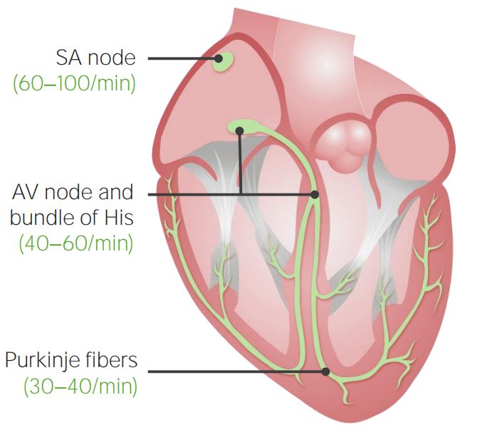
00:01 So we've seen all the individual components of the heart which are very beautiful and very interesting in their own right. 00:08 But just like if you have the world's best violinist and the best cellist, and the best piano player, if they all just start playing on their own at their own tempo, it's gonna sound terrible. 00:18 Same thing with the heart, just like the orchestra needs a conductor to get everything flowing together in a coordinated fashion. 00:25 The heart needs a conduction system. 00:28 And so that's what's going to help make sure that the heart beats in an orderly fashion every time from beat to beat. 00:34 But before we talk about the conduction system, and how electrical impulses do travel, We have to point out where they can't. 00:43 And there's some important essentially insulation of the heart, formed by these collagenist networks called the cardiac or fibrous skeleton that exist between the atria and the ventricles. 00:56 Okay, so where do these pathways actually go then? Well, all conduction starts at the sinoatrial or SA node, sitting way back at the top of the right atrium. 01:10 And then the conduction has to go through the atria. 01:14 And there are various things called internodal tracts, that conduction travels along to reach the next node, hence, the term internodal. 01:23 And we have anterior internodal tracts in the right atrium. 01:27 We have a Bachmann bundle over on the left. 01:31 We have a tract in the middle of the right atrium, sometimes called a Wenckebach tract. 01:36 And then a posterior pathway in the right atrium, sometimes called the Thorel tract. 01:41 The important thing, though, is that all of these internodal tracts are pathways to go from the SA node all the way to the next point, which is the AV node. 01:53 And this atrioventricular node is where everything can kind of take a breath, gather itself, and then go on together into the ventricles in a coordinated fashion. 02:05 And it can do so because of that fibrous skeleton not allowing any pathways to jump across to the ventricle early. 02:12 Everything has to meet up and go through this AV node. 02:17 And then when everything's ready to go with the AV node, it will pierce that fiber skeleton at something called the the bundle of His. 02:24 And ideally, that's the only way for conduction to travel. 02:29 Then it can reach the ventricular septum and branch on either side of it to form a left bundle branch and a right bundle branch. 02:37 So let's look at where these nodes are located. 02:40 Starting with the SA node. 02:43 To find it, we find the superior vena cava right as it's about to join the right atrium. 02:49 And that landmark we saw a little while ago called the Crista terminalis that is the border between the smooth and the bumpy part essentially between the atrial appendage and the rest of the atrium is our border. 03:01 It's right before we hit that Crista terminalis. 03:04 That we're going to find the SA node. 03:08 So how are we going to find the AV node though? That's going to be a little bit more complicated and require finding Koch's triangle. 03:17 So what is Koch's triangle? Well, one side of it is the opening of our coronary sinus. 03:24 To find the other side, we have to look how that valve in front of the coronary sinus and the IVC continue upward as something called the Tendon of Todaro. 03:36 And then we're going to look at the tricuspid valve where it meets the heart. We call that the anulus. 03:43 And we have three sides that vaguely form a triangle that points to a very tiny thin portion of the ventricular septum called the membranous septum. 03:54 It's called the membranous septum because it's just a thin membrane of connective tissue, as opposed to a thick wall of heart muscle like the rest of the septum. 04:03 And it's within this triangle that will find the AV node. 04:09 And right after the AV node is where the fiber skeleton is. 04:13 And that's where the bundle of His is going to penetrate through to reach the ventricular septum, and branch into our left and right bundle branches on either side of the septum. 04:27 So let's look at it from the right side. 04:29 We have our bundle of His penetrating through that insulated layer of the skeleton of the cardiac skeleton. 04:37 Giving rise to our left bundle branch and disappearing from our point of view, because it's going over to the right. 04:43 And then on this side, we have the right bundle branch coming down along the right side of the ventricular septum. 04:53 And then, we have this weird little quirk of the right side called the Septomarginal trabecula or the moderator band. 04:59 That receives a little branch going over to the anterior papillary muscle. 05:04 And that's to make sure that papillary muscle, that's kind of far away from the others will contract at the right time. 05:11 Otherwise, these bundle branches on either side will finally terminate into the free walls at something called the Purkinje network to help stimulate all of these heart muscle cells to contract at the right time. 05:25 So to summarize, we have the SA node, generating the impulse, traveling through the atria via these inter nodal tracts to reach the AV node. 05:36 And then only through the bundle of His can it reach the ventricular septum, as left and right bundle branches, And then eventually out to the rest of the heart via the Purkinje fibers. 05:50 Now, the SA node has its own automaticity. 05:54 But there are nerves that innervate the heart. 05:57 And so, these nerves can influence how the heart beats just not really tell it exactly how. 06:04 So, what do I mean. Well, hormones and nerves such as the vagus nerve, and the sympathetic trunk will form a plexus called the cardiac plexus that can provide autonomic innervation to the heart that may raise or lower the heartbeat. 06:22 For example, parasympathetic lowers, sympathetic raises. 06:24 Just like hormones can do the same thing, like epinephrine will raise the heartbeat. 06:29 But it's important to remember that all the conduction system, those aren't nerves, they're just heart muscle cells modified for conduction instead of contraction. 06:38 And they're automatic, the SA node, as long as it has a blood supply, it will just keep beating away at about 100 beats per minute. 06:46 Fact, if you've ever seen an a heart transplant, the heart being removed from the body and it continues to beat, that shows you that the SA node has its own automaticity. 06:54 It has its own pacemaker cells. 06:57 So this innervation really just modifies the SA node. 07:01 It doesn't really tell it what to do.
About the Lecture
The lecture Cardiac Conduction System by Darren Salmi, MD, MS is from the course Thorax Anatomy.
Included Quiz Questions
Which structure usually functions as the pacemaker, initiating the cardiac conduction cycle?
- Sinoatrial node
- Node of Ranvier
- Atrioventricular node
- Purkinje fibers
- Right atrial appendage
Which of the following is NOT a conduction pathway from the SA to the AV node?
- Superficial internodal tract
- Wenckebach tract
- Thorel tract
- Anterior internodal tract
Which landmark is not associated with the AV node?
- Crista terminalis
- Tendon of Todaro
- Tricuspid annulus
- Opening of the coronary sinus
- Membranous ventricular septum
What transmits electrical signals from the AV node to the ventricular septum?
- Bundle of His
- Purkinje fibers
- Moderator band
- SA node
- Crista terminalis
Author of lecture Cardiac Conduction System

Darren Salmi, MD, MS
Customer reviews
5,0 of 5 stars
| 5 Stars |
|
5 |
| 4 Stars |
|
0 |
| 3 Stars |
|
0 |
| 2 Stars |
|
0 |
| 1 Star |
|
0 |
