Playlist
Show Playlist
Hide Playlist
Anatomy of the Ureters
-
Slides Anatomy of the Ureters.pdf
-
Download Lecture Overview
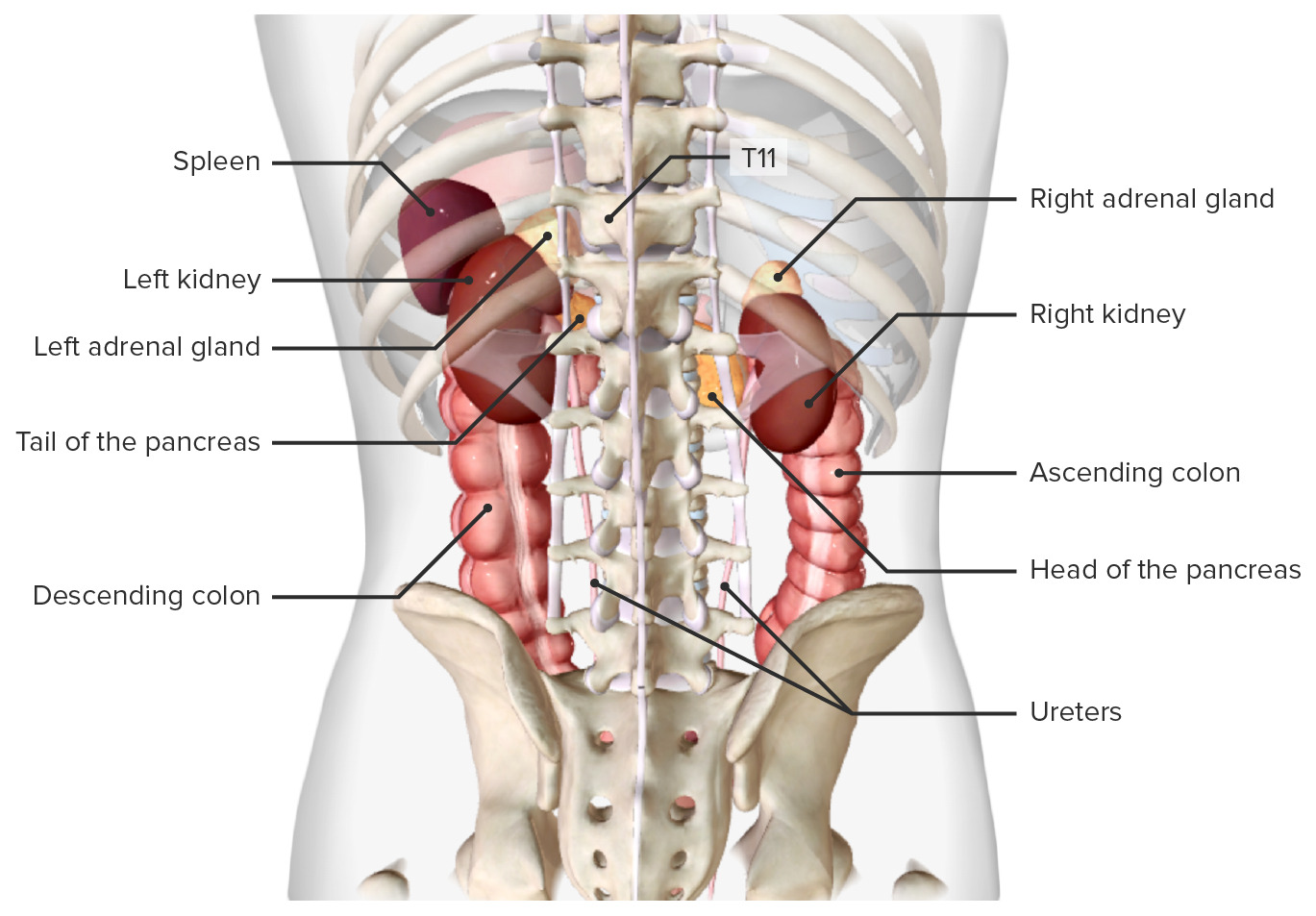
00:01 So now let's have a look at the ureters. 00:03 The ureters are very small channels, tubes that pass away from each of the kidney. 00:10 Obviously, we have two of them, and they're important ureters. 00:14 They have peristaltic contractions within them. 00:17 So they're made of smooth muscle, and they're controlled by the autonomic nervous system. 00:22 The ureters come away from the kidney, and they pass inferiorly along the posterior abdominal wall, and they're destined for the bladder. 00:30 Here we can see they're around 25 to 30 centimeters, but obviously, this will depend on the height of the individual. 00:38 The ureters, as I said, pass along the posterior abdominal wall, and there's some important landmarks we need to be familiar with. 00:45 Running down the posterior abdominal wall to supply the gonads, either the testes or the ovaries in the male or the female, we have the gonadal vessels, so the grown adult artery and vein. 00:56 The ureters run alongside these structures. 00:59 They also run alongside the genital femoral nerve, which we spoke about previously, when we looked at the inguinal canal region. 01:05 The genital femoral nerve is running alongside the anterior surface of psoas major heading for the deep inguinal ring. 01:12 Along its journey, it all run alongside the ureter. 01:16 We also see it runs anterior to the common ilaac vessels, so the iliac vessels, the iliac artery. 01:23 The common iliac artery comes off the bifurcation of the aorta. 01:28 The common iliac veins unite to form the inferior vena cava. 01:32 And as these blood vessels are positioned on the lateral margin of the lesser pelvis, we can see the ureter is running anterior to them. 01:42 Along this course of descending all the way down to the bony pelvis and merging with the bladder, there are some constrictions where actually the tube of the ureter narrows. 01:53 There's a couple of these. 01:54 The first is the pelvico-ureteric junction. 01:58 That's where the renal pelvis starts to narrow down into the ureter itself. 02:04 As the ureter then passes over the common iliac blood vessels, we can see that there's actually a potential sigh of constriction here. 02:12 As it's laying directly flat on these blood vessels, there's a potential constriction. 02:17 We also see the final constriction is actually as the ureters pass into the wall of the bladder. 02:23 These sites are really important because if he were to have a renal stone that situated within the kidney, and it passes all the way down through the ureters, then this renal calculus can actually be obstructed at one of these sites. 02:40 Let's have a very brief look at the arterial supply to the ureter. 02:44 So now we can see the ureter again passing away from the kidney. 02:48 And we can see we have the renal artery passing towards the kidney. 02:52 Coming off that renal artery, there will be a branch going to the ureter, and they'll also be a branch going to the ureter coming off the gonadal archery. 03:01 Remember I mentioned, the ureter runs alongside the gonadal artery as it descends down the posterior abdominal wall. 03:07 They'll also be numerous branches quite small, that passed directly from the aorta and from the common iliac artery. 03:14 So as the ureter descends, its blood supply will be picked up by various arteries that are running adjacent to it. 03:22 Be that the renal artery gonadal artery, the aorta itself, or the common iliac internal iliac blood vessels.
About the Lecture
The lecture Anatomy of the Ureters by James Pickering, PhD is from the course Anatomy of the Urinary System and Suprarenal Glands.
Included Quiz Questions
Which statement regarding the ureters is inaccurate?
- They are intraperitoneal throughout their course.
- They are retroperitoneal throughout their course.
- They convey urine into the bladder.
- They are constricted at the pelvic brim.
- They are constricted at their entrance into the bladder.
Which of the following locations is NOT a site of ureter constriction?
- Along the transverse processes of the lumbar vertebrae
- Junction of the ureter and the renal pelvis
- Crossing the pelvic brim
- Entrance to the bladder
Customer reviews
5,0 of 5 stars
| 5 Stars |
|
5 |
| 4 Stars |
|
0 |
| 3 Stars |
|
0 |
| 2 Stars |
|
0 |
| 1 Star |
|
0 |


