Playlist
Show Playlist
Hide Playlist
Anatomy of the Trachea

-
Slides Anatomy of the Trachea.pdf
-
Download Lecture Overview
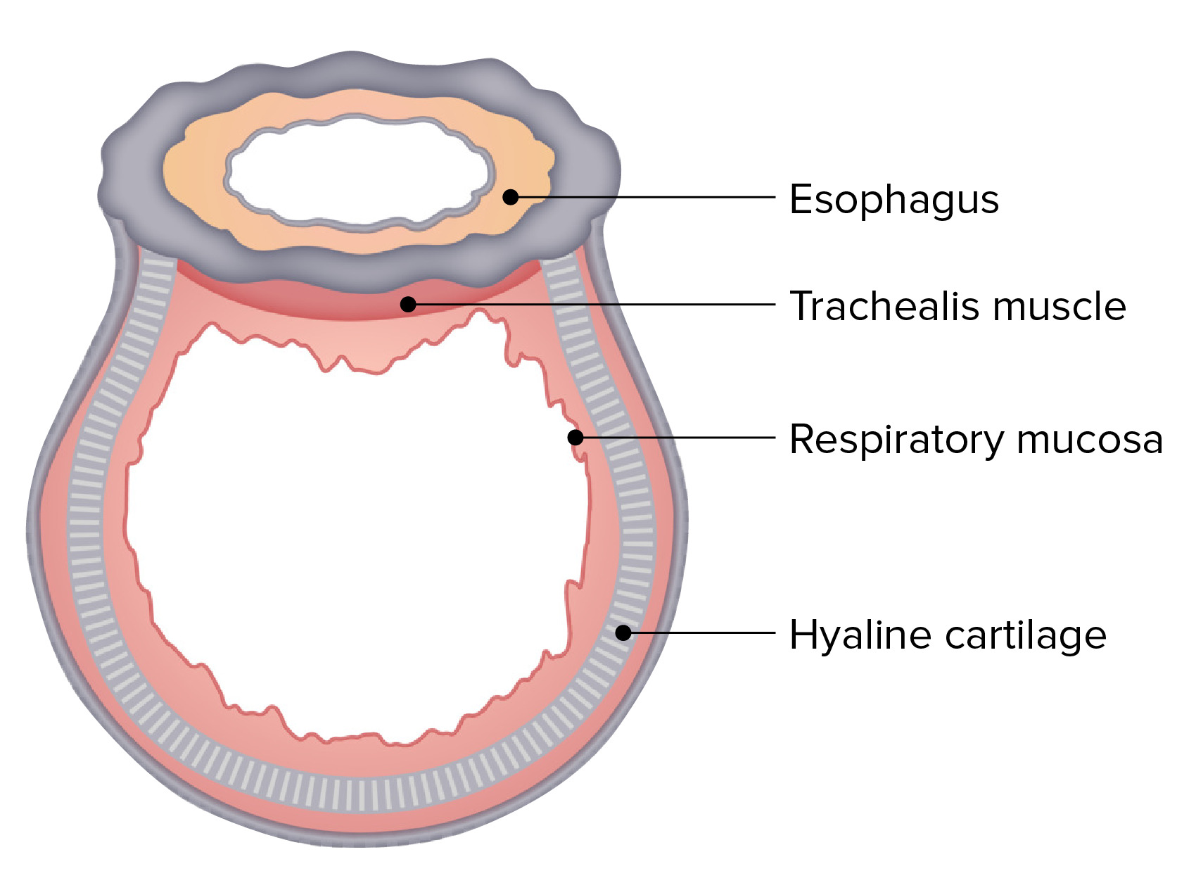
00:01 The next thing we're going to talk about is the trachea. 00:04 Which is not purely a thoracic structure, because it does originate in the neck, and then comes down into the thorax before it bifurcates at a point called the carina into two bronchi. 00:17 A right mainstem bronchus and a left mainstem bronchus. 00:23 And the trachea is made up of about 15 to 20 cartilaginous rings all the way down that helped protect the airway on its way to and from the lungs. 00:32 So, let's look a little closer at this. 00:35 So here we see the trachea, and these cartilaginous rings. 00:40 That we have in quotes here, because they're not rings in the sense that they go all the way around like a circle. 00:47 The cartilage actually ends posterior early, and then those two ends are joined by smooth muscle instead. 00:56 Now, that's pretty useful, for a couple reasons. 01:00 One, that smooth muscle could contract and narrow the lumen when you cough, and by doing so making a narrower lumen makes the air go out faster and better remove any debris or anything that got caught up in the trachea. 01:15 The other advantage is that just posterior to the trachea is the esophagus. 01:22 And this way, if you're swallowing a bolus of food, instead of rubbing up against a firm and movable cartilage, you have a little bit of give up against this smooth muscle. 01:32 And they're very closely aligned. And that's not a coincidence, it's because they develop very similarly embryologically. 01:42 So, the parts of the trachea. 01:45 As we mentioned, the trachea doesn't exist only in the thorax. 01:49 In fact, there's a quite a bit that's in the cervical area. 01:52 So we call that the cervical part where it starts just after the end of the larynx, which is the cricoid cartilage, the last bit of cartilage of the larynx, roughly about the C6 vertebra. 02:07 And then it goes down to reach the superior thoracic aperture. 02:11 And that's where it will become the thoracic part. 02:15 And then it will continue down to the bifurcation until about the T5 vertebra where the carina is in this that's the point where it splits into the bronchi. 02:27 Let's go back and look at this entire tracheal bronchial tree here. 02:31 So it comes down and then branches into a right main and then a left main bronchus. 02:39 And you'll notice there's a bit of a symmetry here. 02:42 We have the right main bronchus being quite a bit more vertically oriented and shorter than the left main bronchus. 02:50 It's also a bit wider. 02:54 The left main bronchus is a bit narrower, and quite a bit longer. 02:59 And it's definitely more horizontal than the right. 03:04 And we can explain this asymmetry by something we'll learn later, which is the heart because the heart doesn't sit right in the center of the thorax, it's somewhat off to the left. 03:12 So the left main has to deal with the presence of the heart. 03:17 This is also why if someone were to swallow or inhale actually a foreign body what we can say aspirate. 03:24 and it went down into the trachea. 03:27 It's going to be more likely to go into the right main bronchus than the left because of this more vertical orientation. 03:35 Now, each mainstem bronchus is going to branch further. 03:40 The right main bronchus is going to branch into secondary or lobar bronchi, namely the right superior, right inferior, and right middle bronchi. 03:51 Whereas on the left, it's going to branch into the left superior low bar and left inferior low bar because there's no middle lobe on the left side. 04:02 Those low bar branches are going to branch again into tertiary or segmental bronchi - 10 on the right, 8 on the left, which are going to branch again into sub segmental bronchi, Eventually down into bronchioles and eventually down to the level of alveoli. 04:19 And that's going to be where gas exchange takes place. 04:22 Let's look at some of the relationships that the trachea has to surrounding features, starting with some of the bones. 04:29 So anteriorly, we see we have that manubrium of the sternum. 04:34 And there was that feature we pointed out that little notch there called the jugular notch. 04:39 And if you very gently put your finger there you might be able to palpate your trachea. 04:45 If we remove the sternum so we can get a little deeper view. 04:48 We can see the arch of the aorta sitting here and it's giving rise to a brachiocephalic trunk crossing anteriorly to the trachea. 04:58 Whereas, the left common carotid artery is coming up to the left of it. 05:03 In terms of venous structures, we see the superior vena cava off to the right of the trachea. 05:09 And it's receiving vein, the left brachiocephalic vein crossing the midline crossing over the trachea anteriorly. 05:18 We already mentioned the esophagus. 05:20 The esophagus is running directly posterior to the trachea for the entirety of its length. 05:26 And that's actually because the trachea embryologically came off of the primitive esophagus. 05:32 So that relationship has always been a very close one. 05:36 If we look laterally from the right, we of course have the right lung and pleura. 05:42 And if we remove that, we can see some more venous structures sitting to the right of the trachea. 05:48 We have the right brachiocephalic vein going down into the superior vena cava, as well as the azygos vein. 05:56 We have some nerves. We have the right vagus nerve going down to the right side of the trachea. 06:01 We also have on the left, the same idea, we have the lung occupying the majority of the lateral surface that we're going to have to remove to see some other structures. 06:11 Namely, this time the arch of the aorta. 06:14 So, we have more arterial stuff on the left, more venous stuff on the right. 06:19 And we see the left subclavian artery here, and we have the left common carotid artery to the left of the trachea. 06:26 And then finally we have the vagus nerve on the left side. 06:30 We also, only on the left, are we able to see a recurrent branch, a recurrent laryngeal branch coming off of the left vagus because this is where the aortic arch is in there's some asymmetry that makes this branching different than it is on the right. 06:49 Now, let's move on to the blood supply of the trachea. 06:52 And in terms of the cervical trachea, the blood supply is coming from the inferior thyroid arteries and veins. 06:59 And that actually makes sense if you've done head neck anatomy, because the thyroid gland actually sits on the upper tracheal rings. 07:08 As for the lower or more thoracic portion of the trachea, we have to look down here at the descending aorta coming off of the arch of the aorta. 07:18 Because there are some branches that come directly off of the descending aorta in this area and those are the bronchial arteries. 07:26 As their name implies, they're also going to supply the bronchi. 07:31 Similarly, the venous drainage in this area comes from what's called the as Azygos system of veins. 07:38 On the left side, we have the Hemiazygos and Accesory Hemiazygos, and they drain over to the Azygos, which in turn goes into the superior vena cava. 07:49 And it's the bronchial veins in this area that will drain into the azygos system. 07:55 On the left side, they'll go into the accessory hemiazygos and the hemiazygos. 08:00 And on the right side, they go directly into the azygos. 08:04 Now, some innervation. 08:06 The main nerve in this area is the vagus nerve. 08:09 So, on the right, the vagus nerve will come down just past the right subclavian artery, and then give a branch called the right recurrent laryngeal nerve that will wrap around and go back towards the trachea on its way up to the larynx. 08:27 However, because of the asymmetry of the aorta here, on the left side, the left vagus nerve comes all the way down to the arch of the aorta. 08:35 And that's where the recurrent laryngeal nerve will branch off, wrapped around the arch of the aorta and go posteriorly towards the trachea on its way back up to the larynx. 08:46 We also have the sympathetic trunk and it's forming some plexuses with these vagus nerves called the pulmonary plexus to provide some autonomic innervation as well.
About the Lecture
The lecture Anatomy of the Trachea by Darren Salmi, MD, MS is from the course Thorax Anatomy.
Included Quiz Questions
What forms the posterior part of the trachea?
- Smooth muscle
- Cartilage
- Bone
- Mucosa
- Ligament
What denotes the most superior part of the trachea?
- Cricoid cartilage
- Thyroid cartilage
- Arytenoid cartilage
- Pharyngeal constrictor
- Thyroid
What is the approximate location of the carina?
- T5
- T2
- T7
- T8
- C7
Which statement about the mainstem bronchi is true?
- The right mainstem bronchus is shorter than the left mainstem bronchus.
- The right mainstem bronchus is less vertical than the left mainstem bronchus.
- The right mainstem bronchus is more narrow than the left mainstem bronchus.
- The right mainstem bronchus is more cartilaginous than the left mainstem bronchus.
- Food more often goes into the left mainstem bronchus.
What artery supplies the cervical trachea?
- Inferior thyroid artery
- Superior thyroid artery
- Inferior laryngeal artery
- Superior laryngeal artery
- Superior esophageal artery
Author of lecture Anatomy of the Trachea

Darren Salmi, MD, MS
Customer reviews
5,0 of 5 stars
| 5 Stars |
|
5 |
| 4 Stars |
|
0 |
| 3 Stars |
|
0 |
| 2 Stars |
|
0 |
| 1 Star |
|
0 |
