Playlist
Show Playlist
Hide Playlist
Anatomy of the Renal Capsule
-
Slides Anatomy of the Renal Capsule.pdf
-
Download Lecture Overview
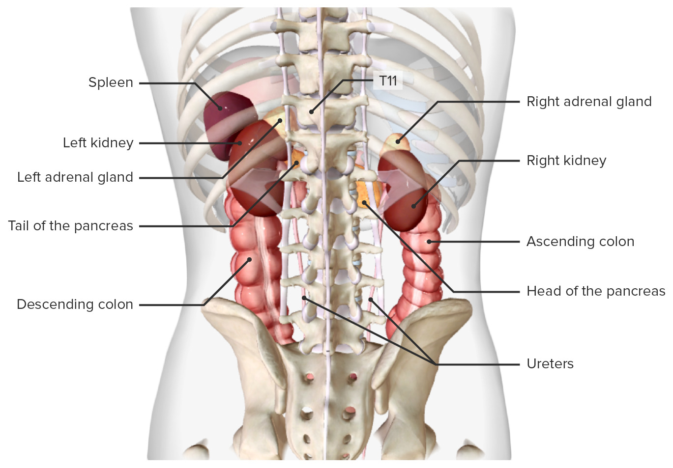
00:01 Now let's carry on looking at the position of the kidney. 00:05 And in this diagram, we're specifically looking at a right kidney. 00:09 But we're looking at it as if we're looking at the patient's through their feet, and they are laying flat on the bed. 00:15 So here posteriorly is at the bottom of the screen. 00:18 And then towards this left side of the screen, we can see the right anterolateral abdominal wall is curving. 00:25 And we can see those three layers of muscles that formed the anterolateral abdominal wall. 00:30 The section has been taken through the kidney and we can still see the aorta and inferior vena cava. 00:37 But here we can see the kidney has been sectioned. 00:40 Surrounding the kidney we have a large portion of fat and this is known as perinephric fat. 00:46 It helps to protect the kidneys. 00:49 This fat is continuous into a space really a hollowing within the kidney that allows blood vessels to pass in and out and also the ureter to leave the kidney. 00:59 This is known as the renal sinus and perinephric fat can escape into this space. 01:06 Surrounding that perinephric fat and really holding the kidney in position we have the renal fascia. 01:13 This is an anterior sheath that lies in front of the kidney behind the peritoneum. 01:19 We can see we have part of this renal fascia projecting posteriorly as well. 01:24 Here we can see it's closely related to transversalis fascia. 01:27 And we can see around transversalis fascia and posterior to the posterior sheath of the renal fascia. 01:35 We also have this paranephric fat. 01:37 That's really sitting alongside the kidney. 01:40 So two important fatty layers: Perinephric fat that sits around the kidney, and then just situated lateral to it, we have this paranephric fat. 01:50 All of which is kept in place by various layers of tough fascia. 01:54 And the transversalis fascia as well that we can see. 01:59 Obviously, most anterior to the transversalis fascia that we can see is the peritoneum and we've discussed that location previously. 02:07 But this gives an indication as to why the kidneys are retroperitoneal they are posterior to the peritoneum. 02:15 So let's have a look at the anatomical relations of the kidneys. 02:19 They sit either side of the aorta and the inferior vena cava, as we can see here. 02:24 And positioned on each superior pole of the kidney we have a supra-renal or adrenal gland. 02:32 Here on the right kidney, you'll appreciate that directly anterior to it. 02:36 We have the liver, and then we have the right flexure as written there of the colon or the hepatic flexure. 02:43 Where the ascending colon becomes the transverse colon. 02:47 So We have some important relations of this right kidney. 02:50 Finally, and the most inferior pole, we have the right kidney associated with the jejunum, that portion of the small intestine. 02:58 We can also see how the duodenum sits very immediately in relation to the kidney. 03:04 But don't forget the kidney is retroperitoneal and the duodenum at this point is also retroperitoneal. 03:10 And these can come into quite close approximation. 03:13 If we look at the left kidney, again, we have an area for the suprarenal or adrenal glands. 03:18 And we have another number of structures such as the stomach on this left hand side. 03:23 We also have the pancreas which passes over its anterior surface as it's heading towards the spleen. 03:29 And we also have the widespread jejunum that is going again, on this anterior aspect of the kidney. 03:36 We can now include the spleen on this most lateral aspect of the left kidney, we're familiar with the spleen being in the upper left quadrant, and here we can see it associated with the left kidney, and also the descending colon very much at the splenic flexure, where the transverse colon is becoming the descending colon, and we're gonna see that located here. 03:56 If we look at the posterior aspect, we can see we have the ribs running along the posterior aspect of the abdominal wall. 04:03 And again, we can see the aorta and the inferior vena cava. 04:07 Here we've indicated the numbers of those ribs being the 11th and 12th ribs that do serve to protect it. 04:13 Running in between those ribs and the kidney, we would have the diaphragm that is running down the posterior abdominal wall coming from the dome that separates the thorax and the abdomen. 04:24 We can also see the transversus abdominus muscle that we spoke about previously. 04:28 And we can see quadratus lumborum muscle here as well. 04:31 We'll also have the psoas major muscle, those muscles that sit on the kidney bed, the actual posterior abdominal wall musculature where the kidneys reside. 04:40 We can also see some important landmarks as in the blood vessels and nerves that are leaving either the aorta, passing into the inferior vena cava, or as we can also see here, some of the nerves that are radiating within this space from the spinal cord. 04:56 We can see the subcostal vessels that are passing underneath the inferior border of the 12th rib. 05:01 Here we can see the subcostal artery and vein and we can also see the iliohypogastric and ilioinguinal nerves passing away from the spinal cord around L1 region and they run between transverse abdominus muscle and internal oblique remember passing around the anterior lateral abdominal wall.
About the Lecture
The lecture Anatomy of the Renal Capsule by James Pickering, PhD is from the course Anatomy of the Urinary System and Suprarenal Glands.
Included Quiz Questions
Which of the following nerves is not found directly posterior to the kidney?
- Genitofemoral
- Subcostal
- Iliohypogastric
- Ilioinguinal
Which statement concerning the renal vasculature is inaccurate?
- The right renal vein is longer than the left.
- The renal veins drain into the inferior vena cava.
- The left gonadal vein drains into the left renal vein.
- The renal veins are positioned anterior to the renal arteries.
Which structure lies most immediately inside the renal fascia?
- Perinephric fat
- Renal cortex
- Paranephric fat
- Peritoneum
- Transversalis fascia
Customer reviews
5,0 of 5 stars
| 5 Stars |
|
5 |
| 4 Stars |
|
0 |
| 3 Stars |
|
0 |
| 2 Stars |
|
0 |
| 1 Star |
|
0 |


