Playlist
Show Playlist
Hide Playlist
Anatomy of the Esophagus

-
Slides Anatomy of the Esophagus.pdf
-
Download Lecture Overview
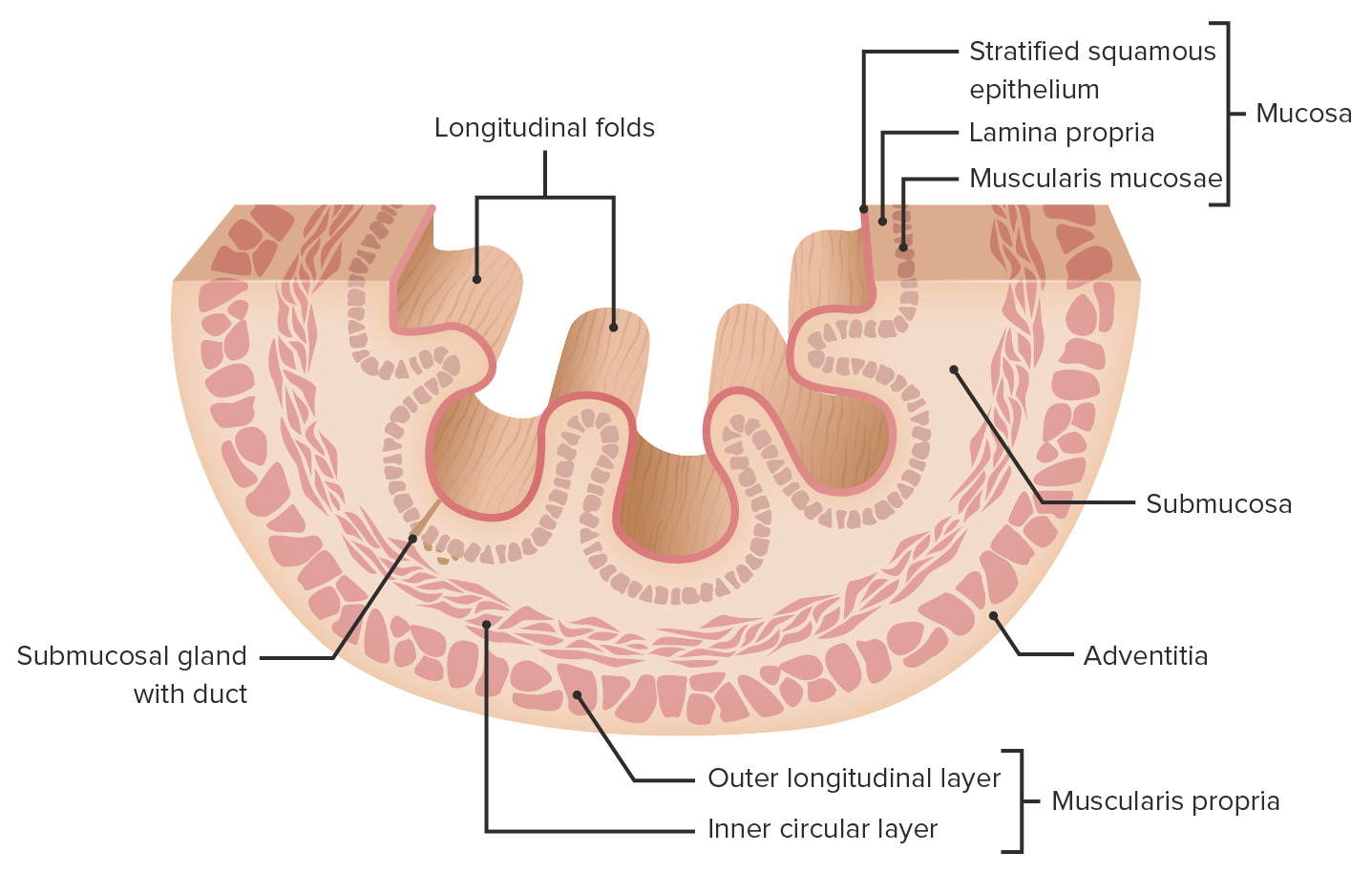
00:01 Now we're going to move on to the esophagus. 00:05 The esophagus is the long muscular tube that connects the pharynx to the stomach. 00:11 And it doesn't just have thoracic parts. 00:14 It starts in the neck and ends in the abdomen. 00:17 It's about 22 to 25 centimeters long. 00:21 And, like the trachea starts around the C6 vertebra, just after the pharynx at something called the pharyngoesophageal junction. 00:32 And it will terminate distally at around the T11 vertebra, where it joins the stomach at the gastroesophageal junction. 00:43 So let's look at the esophagus in relation to some other structures here. 00:47 So here we have the esophagus. 00:49 And we see that a certain point it's going to have to go through the diaphragm. 00:54 And we saw in the diaphragm lecture that there's an opening called the esophageal hiatus that allows the esophagus to travel through the diaphragm and briefly become an abdominal portion before joining the stomach. 01:09 So let's look at those parts. 01:10 So we start off in the neck. 01:13 And so we call that the cervical part. 01:16 And it will terminate right about the area we reach the thorax. 01:20 So right about at the jugular suprasternal notch. 01:24 And there becomes the thoracic part until we hit the diaphragm at the esophageal hiatus. 01:31 And then briefly, we have the abdominal part before entering into the stomach. 01:37 Now, the esophagus is going to do its job by moving food via involuntary contractions called peristalsis. 01:45 However, most of the time nothing's being swallowed. 01:48 So it's going to be closed off by two sphincters, one proximally, and one distally. 01:54 Proximally, is going to be the superior sphincter at that pharyngeal esophageal junction. 02:00 And it's also called the upper esophageal sphincter or UES. 02:05 Distally, we're going to have the inferior sphincter at the gastroesophageal junction. 02:11 And that's often called the lower esophageal sphincter or LES. 02:17 And it's really important for the sphincters to open and close only when they're supposed to. 02:21 So for example, here we see the esophagus coming down to the stomach and the LES the smooth muscle in this area should open and close only with swallowing. 02:32 However, if for some reason, the lower esophageal sphincter can't relax and open up in something called achalasia, any food or liquids that are swallowed can't pass into the stomach. 02:46 And that will cause the esophagus to start to dilate. 02:50 Conversely, closure of that lower esophageal sphincter also serves to keep acid in the stomach where it belongs. 03:00 If it can't contract and it stays open too much, then that acid can go back up into the esophagus, or reflux. 03:09 And this is something very common called gastroesophageal reflux disease. 03:14 And because the esophagus isn't really meant to handle acid like the stomach is, it can cause damage that over time can cause some pretty severe consequences. 03:24 Just because the sphincters open and close when they're supposed to doesn't mean that it's smooth sailing for food the rest of the way. 03:32 There are certain points along the esophagus, where that tube can get a little bit narrower. 03:38 And we call these areas constrictions. 03:41 And so if a large solid object were to be swallowed, it's going to be more likely to lodge at these areas of constriction. 03:51 The first one is the pharyngoesophageal constriction. 03:55 And that's a constriction because of the nearby cartilage, the cricoid cartilage. 04:00 Because that's a pretty intense cartilage, it goes all the way around and forms a complete circle just in front of the esophagus. 04:09 Then we have the thoracic ones. 04:12 And these constructions are going to be caused by the aortic arch and the left bronchus. 04:20 And then finally inferiorly, we have the diaphragmatic constriction, being caused by the presence of the diaphragm at that esophageal hiatus. 04:30 Now let's look at how the esophagus relates to some other structures starting with what sits anterior to it. 04:36 Well, in the trachea section, we mentioned that the trachea and esophagus run together. 04:41 And the trachea indeed sits just anterior to the esophagus because it actually developed from the primitive esophagus very early in development. 04:51 At a certain point, it's going to bifurcate and go into the lungs. 04:55 So then after that point, it's going to be bounded anteriorly by the pericardium. 05:01 And then finally, it's going to hit the diaphragm. 05:05 Posteriorly, it's going to be bounded by the vertebral column all along its length. 05:11 Laterally, on the right side, it's going to have the right parietal pleura, and the azygos vein. 05:19 And on the left, it's going to have the left parietal pleura and some arteries. 05:25 It's going to have the left subclavian artery. 05:27 It's going to have the aortic arch and the descending aorta to its left. 05:33 So what about the blood supply to the esophagus itself? Well, in the cervical esophagus, we have the inferior thyroid artery. 05:43 For the thoracic part, it's coming from branches off of the aorta. 05:50 But when we get to the abdominal part, which is pretty short, it's getting some esophageal branches of the left gastric artery, which is actually a branch of the celiac trunk down in the abdominal aorta. 06:03 Similarly, the venous drainage mirrors the arterial supply so that it's drained in the cervical region by inferior thyroid vein in the thoracic part by the azygos. 06:13 And then, abdominally, by the left gastric vein. 06:16 In terms of innervation, there is a plexus that sits all along the surface of the esophagus, composed of parasympathetic and sympathetic nerves of the autonomic nervous system. 06:29 Where the parasympathetic portion is coming from the vagus nerves. 06:33 And the sympathetic portion is coming from the sympathetic trunk that runs up and down on either side of the vertebral column.
About the Lecture
The lecture Anatomy of the Esophagus by Darren Salmi, MD, MS is from the course Thorax Anatomy.
Included Quiz Questions
What is the approximate length of the esophagus?
- 25 cm
- 20 cm
- 15 cm
- 10 cm
- 17 cm
What is the inferior border of the esophagus?
- T11
- T9
- T7
- T4
- L1
Which abnormality leads to achalasia?
- Failure of relaxation of the lower esophageal sphincter
- Failure of relaxation of the upper esophageal sphincter
- Failure of contraction of the upper esophageal sphincter
- Failure of contraction of the lower esophageal sphincter
- Esophageal metaplasia
What structure is associated with pharyngoesophageal constriction?
- Cricoid cartilage
- Aortic arch
- Epiglottis
- Bronchus
- Thyroid cartilage
Author of lecture Anatomy of the Esophagus

Darren Salmi, MD, MS
Customer reviews
5,0 of 5 stars
| 5 Stars |
|
5 |
| 4 Stars |
|
0 |
| 3 Stars |
|
0 |
| 2 Stars |
|
0 |
| 1 Star |
|
0 |
