Playlist
Show Playlist
Hide Playlist
Anatomy of the Hip

-
Slides Osteopathic Evaluation of the Hip.pdf
-
Download Lecture Overview
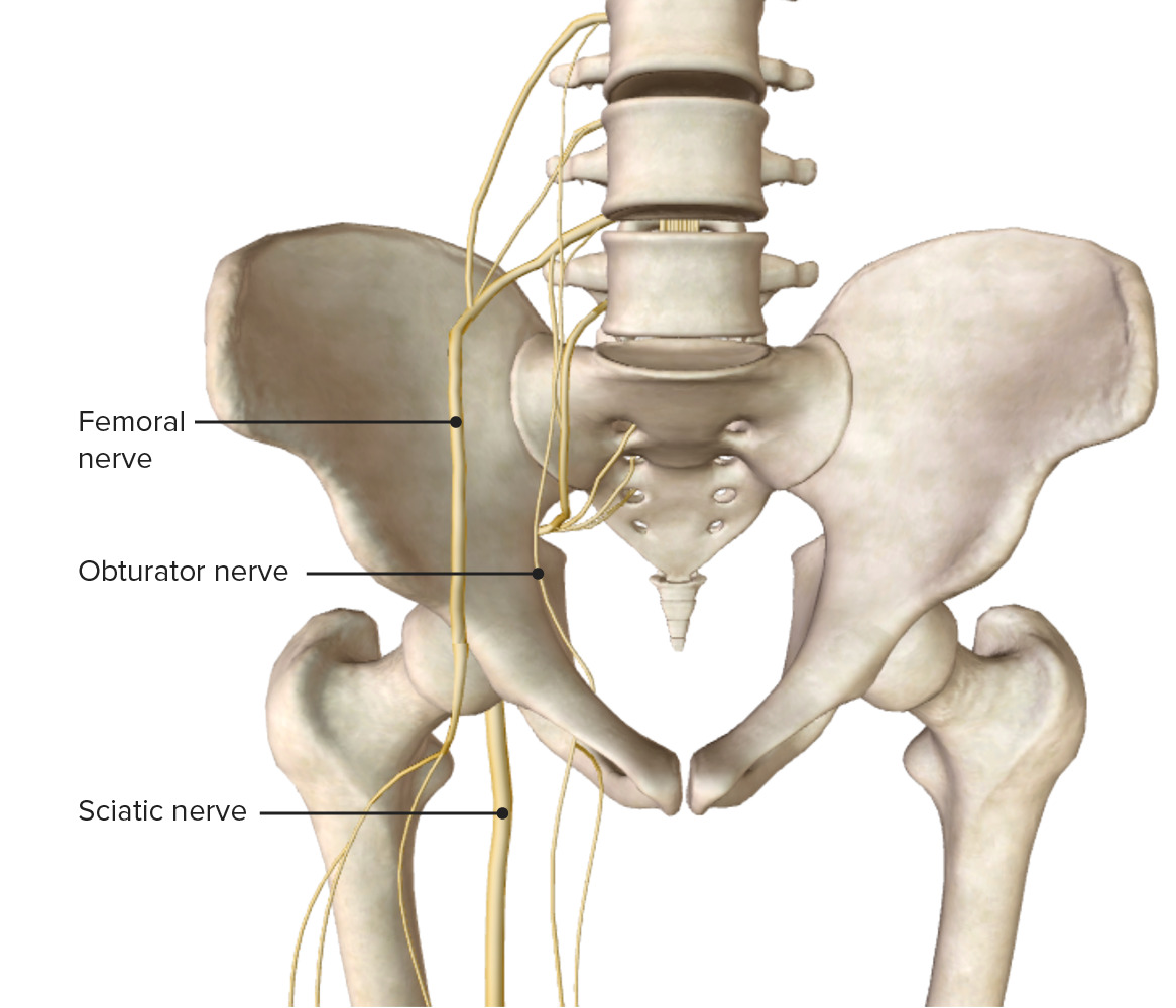
00:01 Osteopathic Evaluation of the Hip Joint Our hip joint is vital for ambulation and locomotion. 00:08 And so patients with hip pathologies and problems will come in and we need to understand the anatomy, physiology, and how to evaluate the joint in order to help better treat our patients with hip problems. 00:22 The hip joint itself consists of 2 bones. 00:25 It’s the articulation between the innominate and the femur. 00:29 So taking a closer look at the femur itself, there is a femoral head, a neck, a greater and lesser trochanter, a shaft, and also a lateral and medial condyle which articulates with the knee. 00:46 The hip joint itself is a ball-and-socket joint and so the surface of the hip articulates in the acetabulum. 00:56 And it’s really deep and provides more stability and is adaptive for high loads. 01:01 This is relatively different compared to the shoulder where the glenoid fossa is relatively shallow and it’s also a ball-and-socket but is not as deep. 01:13 So the hip joint, having more of a deep socket, helps to be more stable but does give it less range of motion compared to the our shoulder joint. 01:22 The capsule extends from the femur to the acetabulum and each part of the innominate. 01:28 The ilium, ischium, and the pubis meet at the acetabulum and forms the articulation of the hip joint with the femur. 01:39 There are many different ligaments that help to stabilize the hip joint itself to keep the femoral head to the acetabulum. 01:48 So anteriorly there’s an iliofemoral and pubofemoral ligament and posteriorly there’s ischiofemoral ligaments. 01:56 And these ligaments make the capsule to help stabilize and keep the femur attached to the acetabulum. 02:07 There are 6 major muscle groups surrounding the hip joint itself. 02:11 We have our flexor and extensors, abductor and adductors, and internal/external rotators. 02:17 So these muscles are large muscles. 02:20 They help to provide motion and movement at the hip joint. 02:25 So let’s have a closer look at the different muscle groups. 02:29 Our hip flexors tend to lie more on the anterior portion of our hip, and so, the iliopsoas is the primary hip flexor. 02:38 It attaches from the anterior bodies of the lumbar spine, blends with the iliacus surrounding the innominate, and attaches into the lesser trochanter of the femur. 02:51 And so when that muscle, the iliopsoas, is injured or spasmodically contracted, that could cause a lot of hip pain. 02:59 And it could also cause the hip to be stuck more in flexion because it attaches to the spine that could prevent someone from straightening out or the pain is worse when they try to straighten out their back. 03:10 The rectus femoris, sartorius, pectineus, tensor fascia latae, and also the gracilis help to make up the primary flexors of the hip. 03:21 The hip extensors tend to lie on the posterior aspect of the hip. 03:26 And here you have your gluteus maximus, your hamstring muscles which include the biceps femoris, the semitendinosus, and semimembranosus, and also the adductor magnus. 03:40 Your hip abductors include your gluteus medius, gluteus minimus, tensor fascia latae, and piriformis. 03:52 Our hip adductors include the adductor brevis, adductor longus, and the adductor magnus. 04:00 There’s the gracilis, pectineus, and also the obturator externus. 04:07 There’s no true pure internal rotators of the hip. 04:10 They have contributions from the gluteus medius and minimus and also contributions from the adductor magnus and longus which help with internal rotation. 04:21 Our hip external rotators include the piriformis muscle, the superior and inferior gemellus, the internal and external obturators, the quadratus femoris, and the gluteus maximus. 04:39 We need to consider blood supply to the hip because sometimes the hip might be suspect to a decrease in blood supply, such as the femoral head, and that could potentially lead to avascular necrosis. 04:51 So in general, the hip joint does get good blood supply but the femoral head itself tends to have less blood flow. 05:01 There is a retinacular arterial system that runs along the neck of the femur and the femoral head itself does get some perforating branches from the medial and lateral circumflex artery. 05:14 Lymphatics of the lower extremity have to flow upward and pass through the hip. 05:19 The hip region is where we try to assess for any lymphadenopathy or swelling of those lymph nodes whenever there’s infection. 05:27 So if someone had a cellulitis of the lower extremity, or someone had some sort of malignancy or anything that really causes infection that inflames the lymph nodes, what we do is we palpate for those swollen lymph nodes along the inguinal canal along the attachments of the hip. 05:46 This is also an important place to look at osteopathically because any sort of musculoskeletal restrictions at the hip and the fascia around the hip, may decrease lymphatic flow from the extremities. 05:56 So it’s important to treat the hip area in order to allow for optimal lymphatic flow.
About the Lecture
The lecture Anatomy of the Hip by Sheldon C. Yao, DO is from the course Osteopathic Diagnosis of the Hip Region.
Author of lecture Anatomy of the Hip

Sheldon C. Yao, DO
Customer reviews
5,0 of 5 stars
| 5 Stars |
|
1 |
| 4 Stars |
|
0 |
| 3 Stars |
|
0 |
| 2 Stars |
|
0 |
| 1 Star |
|
0 |
A very complete summary to refresh knowledge of the hip. Thanks you!
