Playlist
Show Playlist
Hide Playlist
Pituitary Gland: Structure

-
Slides 04 Human Organ Systems Meyer.pdf
-
Download Lecture Overview
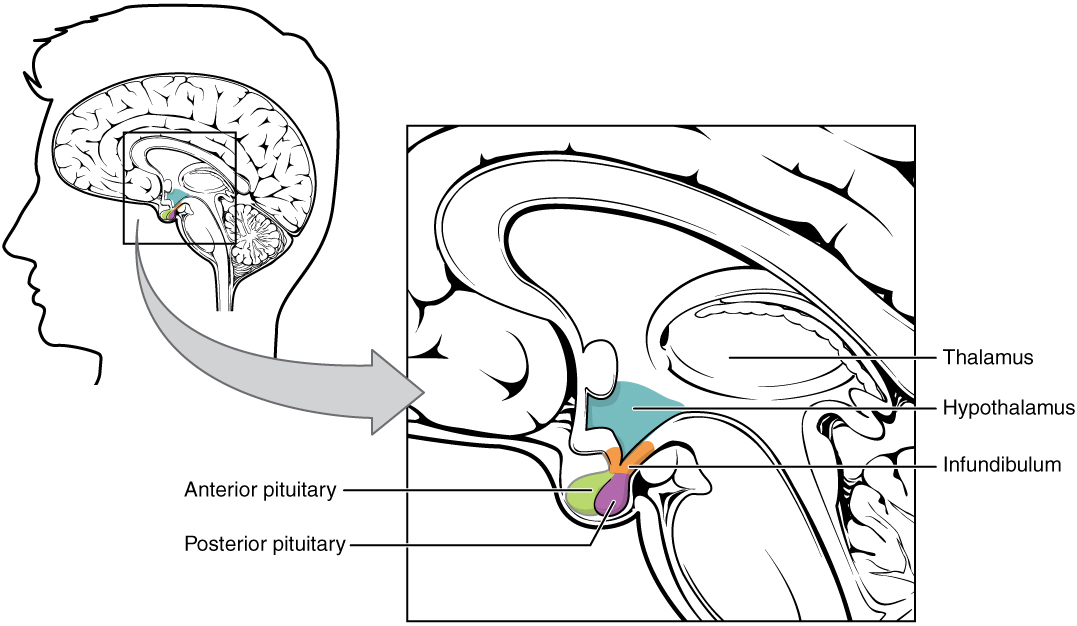
00:01 Well, let’s look at the pituitary gland. It’s often called a hypophysis. Here is, on the left-hand side, a diagram representing the structure of the pituitary gland. And on the right-hand side is a histological section through the pituitary gland. And it’s important that we go through a number of different components. The pituitary gland is really a very small pea-shaped gland. In males, it’s about half a gram in weight. In females that have had two or more children, there can be about one and a half grams. So there’s a difference in size. The pituitary gland sits in a depression called the sella turcica in the sphenoid bone at the base of the brain, at the base of the skull. And as you can see in both these sections, first the section of the image or histological section through the pituitary gland on the right, and the diagram on the left, the pituitary gland is connected to the brain. It’s connected to the hypothalamus through a structure we call the infundibulum. And that’s a very important connection because the hypothalamus influences the secretory nature and products coming out the pituitary gland. Well, the other thing you see in this diagram is that the pituitary gland has two different components, two different major components. On the right-hand side on the histological section, the far left component of that section is a lighter purple color compared to the rather more enlarged component on the right-hand side of this section. 02:03 And this coloration defines or distinguishes the two parts of the pituitary gland. The lighter stained area is the posterior part of the pituitary or the posterior pituitary gland. 02:18 We call it often the pars nervosa or the nervous part or the neurohypophysis because another name for the pituitary gland is the hypophysis. On the right hand side, the darkest stained region represents the anterior pituitary gland, the bit towards the anterior of the body. 02:41 It’s often called the adenohypophysis, adenoma glandula. It’s often called the pars distalis as well. Now, both these components of the pituitary gland are derived from two separate embryological locations. And that’s why they should be really considered two separate glands. The pars nervosa, the nervous component, is derived from a downgrowth from the third ventricle of the brain. So it’s derived from neuroectoderm. The anterior pituitary is derived from an out-pocket of the oropharynx or Rathke's pouch. So it’s derived from oral ectoderm. So these two differences are the reasons why histologically, they are very different when we look at them using a microscope. Now, the diagram on the left-hand side also shows a number of different structures associated with the pituitary gland. There is part of the pars distalis or the anterior lobe of the pituitary, that extends up and wraps around the infundibulum, the part of the stalk or the connection to the hypothalamus. There's another part . They are called the pars intermedia. 04:08 This is of little relevance to us, so I’m not going to cover it in any detail. Have a look at the hypothalamus, that yellow stained component. In the hypothalamus, you can see two nuclei labelled. These nuclei are called the paraventricular nucleus, and also the supraoptic nucleus. They are going to be very very important structures that I’m going to refer to in the next couple of slides. This diagram, again, appears to be rather complicated, but there is only a couple of very simple stories that I want to emphasize. First of all, have a look in the hypothalamus. 04:54 Again, look at the paraventricular nucleus and the supraoptic nucleus. 05:01 A nucleus in the brain is a collection of nerve cell bodies. And they’re shown here on this diagram. Notice though, that the cell bodies project their axons down the infundibulum or the pituitary stalk into the location of the posterior pituitary, the neurohypophysis. 05:25 Now these neurons are neurosecretory. They don’t transmit an impulse. What happens is that oxytocin and ADH, two hormones, are produced by these two nuclei, by the cells in these two nuclei. Oxytocin is produced by mostly a paraventricular nucleus. ADH is produced mostly by the supraoptic nucleus. 05:52 And those secretory products travel down the axons and are stored in axon terminals in the neurohypophysis. And we’ll see that in a later slide. So the secretion of oxytocin and ADH is from neurosecretory cells directly into fenestrated blood capillaries indicated there in the diagram. The blood supply to this region comes from the inferior hypophyseal artery. So that’s the major component of the paraventricular nuclei and the supraoptic nuclei providing secretory products into the posterior pituitary. There’s also another nuclei, group of nuclei that you see in the hypothalamus as well. We’ve got a very long name to it, hypothalamohypophysiotropic nucleus or nuclei. They contain neurons that secrete releasing factors. And those releasing factors get taken up by capillary bed I’ll mention in a moment, up in the hypothalamus. And those releasing factors when they get into the capillary bed, can affect the secretory products from the anterior pituitary. And I’ll go through that in a moment. 07:25 If you look at the adenohypophysis, the anterior pituitary, you’ll notice that in the diagram, there are three cell types represented, basophils, acidophils, and then cells that don’t take up any stain. And this is because in the anterior pituitary, the typical glandular epithelial component of the pituitary gland, there are different cell types based on their staining characteristics. We have cells collectively called chromophils. 08:00 These are the ones that have an affinity for color, and they are the basophils or the acidophils. Then we have the chromophobes which make up a large proportion of the cells that don’t take up the stain. And we’re not quite sure whether this have any functional significance or whether or not they happened to be the chromophils that are just going through a process of resynthesizing their secretory products. 08:28 But I’ll show you these cells in more details in a moment. 08:33 But just imagine now that the anterior pituitary is full of these different secretory cells that secrete different products we’ll see in a moment. And those cells secrete into the vascular system. 08:49 So it’s important now that I explain the vascular supply to the pituitary gland. If we go back to the hypothalamus region, you’ll see just at the top, a vessel called the superior hypophysial artery. Now this artery branches and it provides the hypothalamus with a network of capillaries. It’s called the primary capillary plexus or network. And it’s in just the upper part of the infundibulum, that stalk that connects the pituitary gland to the hypothalamus That’s a very important primary plexus of capillaries because those secretory products, the releasing factors I mentioned earlier from those hypothalamohypophysiotropic nuclei, are secreted into that primary capillary plexus. From that plexus, the blood then flows down through portal veins, along the infundibulum, and then they all supply the anterior pituitary. They don’t go the posterior pituitary. So this is a situation where the releasing factors that are secreted from those nuclei in the hypothalamus, I’m not going to try and say that word again, they pass down through the capillaries and can affect the secretion of the cells in the anterior pituitary. These releasing factors can either stimulate cell secretion from these cells, and they’re very specific what cells they actually stimulate, or they can inhibit secretion from these cells. So there is this control of the secretory products coming out of the anterior pituitary by neurons in the hypothalamus. 10:56 Posterior pituitary, as I said before, does not receive these portal vessels, and therefore, the secretion products coming from the neurosecretory cells in the posterior pituitary are unaffected by these releasing factors. 11:11 This hypothalamic-hypophysial portal system is an extremely important concept to understand and it’s an extremely important piece of histology and microanatomy influencing the function of the anterior pituitary gland. 11:33 Well, here is a little summary of, first of all, on the right-hand side, the components of the pituitary gland, and on the left-hand side, those components in diagrammatic form. And I think it’s important for you to fully understand that there are these components in the hypothalamus, neurons in the hypothalamus, understand where they project their axons and what the function of these projections and their products are in the control of the other glands that we’ll see from these secretory products, and understand also the concept of epithelial cells in the anterior pituitary staining differently and influenced via releasing factors coming down that hypophysial or hypothalamic-hypophysial portal system.
About the Lecture
The lecture Pituitary Gland: Structure by Geoffrey Meyer, PhD is from the course Endocrine Histology.
Included Quiz Questions
The pituitary gland is located in a depression in which of the following bones?
- Sphenoid
- Ethmoid
- Parietal
- Temporal
- Frontal
The posterior pituitary gland is derived from a downgrowth of which of the following brain ventricles?
- Third ventricle
- First ventricle
- Second ventricle
- Fourth ventricle
- Fifth ventricle
The posterior pituitary gland is primarily supplied by which of the following arteries?
- Inferior hypophyseal artery
- Superior hypophyseal artery
- Posterior hypophyseal artery
- Anterior cerebral artery
- Posterior cerebral artery
The anterior pituitary gland is derived from which of the following structures?
- Ectoderm
- Third ventricle
- Endoderm
- Mesoderm
- Fourth ventricle
Which of the following hormones is a posterior pituitary hormone?
- Antidiuretic hormone
- Growth hormone
- Thyroid stimulating hormone
- Luteinizing hormone
- Follicle stimulating hormone
Which of the following hormones is secreted by the paraventricular nucleus of the hypothalamus?
- Oxytocin
- Growth hormone
- Thyroid stimulating hormone
- Adrenocorticotropic hormone
- Prolactin
Author of lecture Pituitary Gland: Structure

Geoffrey Meyer, PhD
Customer reviews
5,0 of 5 stars
| 5 Stars |
|
5 |
| 4 Stars |
|
0 |
| 3 Stars |
|
0 |
| 2 Stars |
|
0 |
| 1 Star |
|
0 |
