Playlist
Show Playlist
Hide Playlist
Alzheimer's Disease: Pathology and Treatment

-
Slides 02 Dementia Neuropathology II.pdf
-
Download Lecture Overview
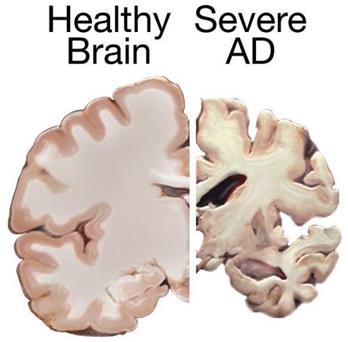
00:01 Gross. What are you gonna find a gross examination? Well, I just told you. You're gonna lose your brain. 00:07 Cortical atrophy, and when you do, what happens to sulci? It gets deeper and deeper and deeper. This is not a good thing. 00:14 You have compensatory ventricular enlargement, in other words, what do we call this? Hydrocephalus ex vacuo, right? Compensatory ventricular enlargement. 00:23 Why? Due to your cortical atrophy. You put these together, don't memorize them. 00:28 It's the same thing, it's just a story. 00:30 Microscopically, what are you gonna find? Okay, now these are interesting. 00:34 What I haven't talked to you about yet in great detail is neurofibrillary tangles. 00:39 But what I have talked to you about and have shown you is the beta-amyloid plaques, correct? So, what did the plaques look like? It looks like a plate. 00:46 So, plate is large. What about neurofibrillary tangle? You know what a sperm looks like? You know what a sperm looks like? It look like little tadpoles, don't they? So, whatever you wanna use here in terms of description. 00:57 Yes, I'm being dramatic because I need you to know the difference, and these two are huge points pathologically on a miscroscopic level. 01:05 So, if you find sperm in the brain, well, do you understand how silly that is? Or tadpoles in the brain, that's your neurofibrillary tangle. 01:13 What is it? It is hyperphosphorylated tau protein. Tau, [ptao]. 01:20 Anyhow, I shouldn't have said [ptao], but tau protein is neurofibrillary tangles. 01:25 These look like tadpoles, there's sperm in the brain. 01:28 Now, let me tell you something. 01:30 Each one of us, as we get older, we're all gonna develop neurofibrillary tangles. 01:34 You can't help it, okay? It's just kinda like as we get older, there's every possibility developing lipofuscin, right? Why? Because we end up losing the ability to properly manage free radicals and such. 01:45 So, neurofibrillary tangle is part of normal aging, however in Alzheimer's disease, there's an accelerated accumulation of hyperphosphorylated tau protein in neurofibrillary tangles. 01:56 So, how much so? What you must understand is that the degree or the concentration or percentage in neurofibrillary tangle, as it increases, correlates with the severity of the disease. Pretty important stuff, isn't it? Oh, yeah. 02:12 Now, do you understand as to why I've been so dramatic on this particular point? Next, so what is it? It's tau, T-A-U. What happened to that tau? It got hyperphosphorylated. Take you back by chemistry real quick, give me the name of the enzyme that causes phosphorylation. 02:31 I'm sorry, what did you say? I think you said kinase. Right on. Exactly right. It's kinase. 02:37 So, you're gonna have a type of kinase that is undergoing or accelerating its phosphorylation of your tau. 02:45 And so therefore, technically speaking, it's axonal microtubule-associated proteins. 02:50 I would have to say, every single word in this particular image or this slide, is of utmost important for you to understand pathologically. 03:00 Every single word. Let's move on. 03:04 Microscopically, what else are you gonna find? Here's the plate. 03:07 These are called senile neuritic plaques. 03:10 You remember that picture that I just showed you where there was green rods? Right? Green rods? That green rod represent -- it's actually quite interesting. It's amyloid. 03:21 It's the amyloid. What kind? What do you mean what kid? Is it kappa lambda? Is it beta-amyloid? Is it -- I don't know, Portuguese? So, all I'm saying is, it's amyloid. 03:37 As far as you're concerned, amyloid, Congo red stain, and then it appears as being your apple green birefringence. 03:43 You know that. So, green is a wonderful color to show you there. 03:45 And so therefore, a bunch of these green rods are beta-amyloid that are now aggregating, and there then form a plaque or a plate. 03:52 Focal, spherical, collection of your dilated, tortuous, silver -- silver staining neuritic processes. 04:01 And so, therefore we call this dystrophic neurites. 04:15 And around, well, that's around the plaque. 04:18 Beta amyloid deposition is within the dysfunctional axonal end or neurite and we call it dystrophic neurites, and in the middle, you'll have your beta amyloid plaque. 04:32 Now the dominant component is going to be your amyloid precursor protein which then breaks down into, and it stands for beta amyloid. Is that clear? Be careful please. It says beta amyloid, so amyloid beta in other words. 04:48 A, representing amyloid, the operative term is beta. 04:53 Where is it coming from? APP. 04:55 What does that stand for? Amyloid precursor protein, once again, yet, another important microscopic feature of Alzheimer's disease. 05:03 What else? Well, found in abundance in the hippocampus. 05:09 What is found in abundance in the hippocampus? Don't just memorize what I'm saying, ask me. 05:14 Dr. Raj, what is found in the hippocampus and your olfactory bulb? What did we just talk about? What are the tadpoles called in your fibrillary tangles, what is it? Hyperphosphorylated tau protein. 05:26 And then what was the other one, that was the beta amyloid collection of plaque -- good, that was the beta amyloid. 05:31 Ah, that is what is found in the hippocampus and olfactory bulb and what happens? Remember that picture that I showed you of the hippocampus? Now your telling -- in Alzheimer's disease, you're gonna tell me, Dr. Raj, I'm sorry but I didn't find the hippocampus in that Alzheimer's disease brain. 05:46 Very good, that's exactly right. At some point in time the hippocampus is gone. 05:51 What about that amyloid? Oh, it's everywhere. Oh, it's everywhere. 05:55 It's wreaking havoc. And where else could accumulate? Around your blood vessels what do you think you call this? Pathology of your blood vessel in the brain, amyloid angiopathy. 06:05 Clear? Good stuff. Hope you're liking it. 06:09 Know everything about Alzheimer's disease and make sure that you medicine all your life, it's more that you keep your brain active. 06:19 Research is showing that perhaps you'd be able to prevent Alzheimer's disease. 06:24 Just saying, let's move on. 06:26 These images are photomicrographs of brain tissue from individuals with Alzheimer disease. The image on the left displays a silver stain that selectively highlights tau protein deposits. 06:39 These deposits manifest as neurofibrillary tangles within the cell bodies of neurons, indicated by a single arrow, and as neuropil threads within the dendrites, indicated by a double arrow. 06:50 On the right, the brain tissue has been stained using a tau-specific immunostaining technique, which reveals a high concentration of tau-positive neuropil threads, denoted by red arrows. 07:02 Additionally, a neuritic plaque is identified by a green arrow, and a neurofibrillary tangle is marked with a blue arrow. 07:09 Very good you all, let's move on. 07:11 Alzheimer's -- so what's my prevention of progression? That kinda difficult, but maybe acetylcholinesterase inhibitors. 07:19 What does that mean to you? How about convenience, A and A? What does that mean? Acetylcholine and Alzheimer's. 07:28 Close your eyes, acetylcholinesterase, where are you? Normally, where are you? Okay you're at the junction, good, and normally responsible for breaking down your acetylcholine. 07:40 And so therefore in the brain, if you are able to inhibit acetylcholinesterase, the drugs include donepezil, that's a big one. 07:48 Another one called rivastigmine, and galantamine Know these and therefore -- what are you gonna do with your levels of acetylcholine? You're gonna increase it and maybe perhaps, by doing so, prevent your progression. 08:00 It is proven to slow the progression; however, it is not the cure, definitely not the cure, unfortunately. 08:06 Additional therapies include memantine, an NMDA receptor antagonist, and recently approved monoclonal antibodies directed against beta amyloid. 08:15 Prophylaxis maybe anti-inflammatory, antioxidants such as Vitamin E, but there's really no data so I'm just gonna quickly go through this. 08:23 As soon as you see the symptoms, oh, boy, yeah, definitely depression, agitation, sleep disorders, late onset hallucinations, delusions -- they have all kinds of issues, don't they? They walk out the door maybe they're naked, they don't know how to come back, you find them wandering. 08:37 That you do, you do find these such cases, don't you? And introduce yourself every single day because they forget as to who you are, and depression, obviously. 08:47 So lots of associated symptoms -- spend a little bit of time with Alzheimer's disease, please. 08:51 Summary: Risk factors -- female, genetic predisposition. I talked to you about PS1 and PS2. 08:57 Specifically talked to you about APOE4, that's your focus should be on. 09:01 They also have Down syndrome and you have Trisomy 21 -- things we've talked about in great detail. 09:06 Preventive medicine -- antioxidants, but really no data out there; continued mental activity -- aha, I told you about that, huh? Hence, my addiction to medicine, a medicine meaning mental medicine. I don't take anything here, uh-uh. Move on. 09:19 Signs and symptoms -- short term memory loss, getting lost in familiar places. 09:23 Differentials -- we'll get into these. Temporary dementia, formerly known as Pick disease. 09:31 What else do we have? Wernicke-Korsakoff -- what do you know about Wernicke-Korsakoff? Probably drinker, alcohol. What made then happen over a period of time? You lose your thiamine, maybe damaged the hippocampus, so on and so forth. 09:43 Normal pressure hydrocephalus, we talked about this and we'll talk about this further, neurosyphilis. 09:49 Diagnostic workup -- Now how do you -- this is an important paragraph. 09:54 You wanna rule things out, okay. 09:57 So you're doing a neurologic examination and you found papilledema and what are you thinking of possibly? So this is not partial hydrocephalus -- Argyll Robertson pupil that, too should indicate neurosyphilis. 10:09 Ophthalmoplegia, here maybe Wernicke's or progressive supranuclear palsy and always check for TSH and B12. 10:19 Diagnostic workup -- all will be to see if you can rule things in, rule things out so that you're on the right track of management. 10:25 Extremely important, in terms of management, let's then move on to the next column. 10:29 Here we have cholinesterase inhibitors and here, where are you? You're trying to save the brain, so this is not so much about Myasthenia Gravis, please don't do that right now. 10:38 I want you to know about donepezil and we talked about rivastigmine and galantamine. 10:43 And side effects are mainly bradycardia and vivid, vivid dreams due to such drugs. Keep this in mind.
About the Lecture
The lecture Alzheimer's Disease: Pathology and Treatment by Carlo Raj, MD is from the course Dementia. It contains the following chapters:
- Alzheimer's Disease: Pathology
- Alzheimer's Disease: Treatment
Included Quiz Questions
Which of the following is most likely seen in a patient with Alzheimer disease?
- Cognitive impairment
- Argyll Robertson pupil
- Papilledema
- Internuclear ophthalmoplegia
- Asterixis
What is the composition of neurofibrillary tangles?
- Paired helical filaments containing hyperphosphorylated tau protein
- Paired helical filaments containing hypophosphorylated tau protein
- Paired helical filaments inside the nucleus
- Paired helical filaments containing hypophosphorylated axonal microtubule-associated protein
- Paired helical filaments inside the cell wall
In Alzheimer's disease, what is the significance of finding amyloid beta in the hippocampus and olfactory bulb?
- It correlates with the severity of the disease.
- It indicates a normal aging process.
- It is a hallmark of early-onset Alzheimer's disease.
- It is a sign of vascular pathology in the brain.
- It suggests the presence of neurosyphilis.
Which medication class is used in Alzheimer's disease to slow the progression by increasing the levels of acetylcholine?
- Acetylcholinesterase inhibitors
- NMDA receptor antagonists
- Dopamine agonists
- Monoamine oxidase inhibitors
- Corticosteroids
What is the stain that can be used to detect neurofibrillary tangles?
- Silver stain
- Congo red stain
- India ink stain
- Sudan Black B stain
- Oil Red O stain
What is the compensatory ventricular enlargement occurring in Alzheimer disease called?
- Hydrocephalus ex-vacuo
- Obstructive hydrocephalus
- Non-obstructive hydrocephalus
- Normal pressure hydrocephalus
- Communicating hydrocephalus
Which of the following is NOT seen in the pathology of Alzheimer disease?
- Hypophosphorylated tau protein
- Hyperphosphorylated tau protein
- Dystrophic neurites
- Amyloid precursor protein
- Neurofibrillary tangles
Which of the following is NOT used in preventing the progression of Alzheimer disease?
- Neostigmine
- Donepezil
- Rivastigmine
- Galantamine
Author of lecture Alzheimer's Disease: Pathology and Treatment

Carlo Raj, MD
Customer reviews
5,0 of 5 stars
| 5 Stars |
|
2 |
| 4 Stars |
|
0 |
| 3 Stars |
|
0 |
| 2 Stars |
|
0 |
| 1 Star |
|
0 |
Great professor! Thank you for your lectures! it is short, informative, and NOT boring!
Excellent explanation in every perceptive ! I would like to have a bit more pictures in the PPT !
