Playlist
Show Playlist
Hide Playlist
Scapulohumeral Muscles – Axilla and Brachial Plexus

-
Slides 04 UpperLimbAnatomy Pickering.pdf
-
Download Lecture Overview
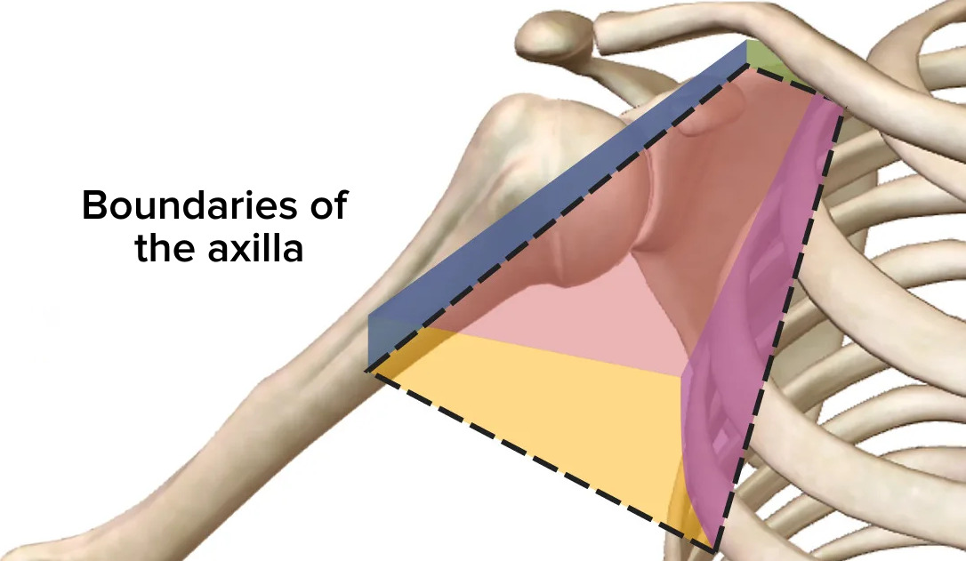
00:01 So let’s look at these spaces a little bit more detail. Here, we’re going to look at the quadrangular space. And the quadrangular space is important because it contains the axillary nerve. The axillary nerve, as we remember, innervates the deltoid muscle and it’s coming away from the brachial plexus, which is located within the axilla. 00:21 So the axillary nerve comes from the brachial plexus within the axilla and it passes out towards deltoid muscle by passing through the quadrangular space. Also coming through this space, we have the posterior humeral circumflex artery and accompanying vein, and these run around the surgical neck of the humerus. So what makes up the boundaries of this quadrangular space? Well, on the screen at the moment, we can see an anterior view of the right shoulder. So we can see the chest wall has been removed and we can see here we’ve got subscapularis muscle, and then down here, we can see we’ve got the humerus and various muscles. We'd remind ourselves that we’ve got teres major muscle running up here. So, what we can see is if we look at the quadrangular space, it’s going to be this muscle in here, the quadrangular space. We’re going to see this muscle in here. 01:22 We can see that inferiorly, we’re going to have a muscle. Inferiorly, we’re going to have a muscle and that is teres major. We can see teres major running here. 01:33 We can also see that if we go medially, we can see we’ve got a muscle here, and this muscle is the long head of triceps. We can see that here. If we look laterally, then we can see we actually got the humerus here, and this is going to be the surgical neck of the humerus. 01:51 Superiorly, teres minor is quite difficult to see actually because subscapularis muscle is in the way. But we’d have teres minor running across in this direction. 02:03 Remember, teres minor comes away from the scapula superior to teres major. So here, we can see this small little space. Superiorly, we’ve had teres minor. Medially, we’d have the long head of triceps brachii. Inferiorly here, we’d have teres major. And laterally, we'd have the surgical neck of the humerus. And this is your quadrangular space. Coming through that space is going to be the axillary nerve and the posterior humeral circumflex artery and vein. If we move on to the next slide, we can actually see if we just remember the quadrangular space. We can pick it up again here. And now, we can see that superior boundaries, teres minor. So in this posterior view of the right shoulder, again, we can see superiorly now we have teres minor; inferiorly, we have teres major; laterally, we have the surgical neck of the humerus; and medially, we have triceps brachii, the long head. And again, we can see the quadrangular space. But this slide, I want to talk about another space, and this space is known as the triangular space. And we can see the triangular space here. The triangular space is important as it allows the circumflex scapula vessels to pass out from the axilla and go to supply the scapula region. It has got three boundaries. That’s why it’s called the triangular space. And those three boundaries are superiorly, teres minor; inferiorly, teres major; and laterally, we’ve now got the long head of triceps again. So we can see that triceps, the long head of triceps is forming the medial boundary of the quadrangular space and it’s forming the lateral boundary of the triangular space; the medial boundary of the quadrangular space, the lateral boundary of the triangular space. With teres minor running across superiorly for both of these spaces, and teres major running inferiorly for both of these spaces. So being aware of the musculature of the axilla, what forms its boundaries, helps us to appreciate the boundaries of these spaces. So this is the triangular space. The final space I want to talk about is known as the triangular interval. 04:35 And this is important as it allows structures to pass out of the axilla and pass to the posterior compartment of the arm to supply the triceps muscles. We have the radial nerve and we have the profunda brachii artery. Now, this is slightly harder to see because the space is actually closed here. But what we can see again is superiorly, we’ve got teres major, and the space is actually going to sit in between the humerus and the triceps long head. So here we can see we've got triceps long head, we've got the humerus, and then superiorly, we've got teres major. And just in this little interval, this little slit here, we find the triangular interval. And coming out of here, we have the radial nerve and the profunda brachii artery. So this is not as clear as the quadrangular and the triangular, but here, just in this little slit here, superiorly, teres major; laterally, the humerus; medially, the long head of triceps brachii, we have the triangular interval. So, very important spaces that allows structures to pass out of the axilla to the wider areas.
About the Lecture
The lecture Scapulohumeral Muscles – Axilla and Brachial Plexus by James Pickering, PhD is from the course Upper Limb Anatomy [Archive].
Included Quiz Questions
Which structure forms the inferior boundary of the quadrangular space?
- Teres major
- Surgical neck of the humerus
- Long head of the triceps
- Teres minor
- Subscapularis
Which structures pass through the quadrangular space?
- Axillary nerve and posterior humeral circumflex artery
- Axillary nerve and axillary artery
- Axillary nerve and axillary vein
- Axillary nerve and anterior humeral circumflex artery
- Axillary artery and vein
Which statement regarding the triangular space is correct?
- The superior boundary is formed by the teres minor.
- The radial nerve passes through it.
- The inferior boundary is formed by the teres minor.
- The lateral boundary is formed by the short head of the triceps.
- The circumflex scapular vessels form the medial border.
Which of the following structures pass through the triangular interval?
- Radial nerve
- Axillary nerve
- Axillary artery
- Brachial artery
- Musculocutaneous nerve
Which structure forms a boundary of the quadrangular (quadrilateral) space?
- Long head of the triceps
- Long head of the biceps
- Short head of the triceps
- Surgical neck of the radius
- Surgical neck of the ulna
Author of lecture Scapulohumeral Muscles – Axilla and Brachial Plexus

James Pickering, PhD
Customer reviews
5,0 of 5 stars
| 5 Stars |
|
1 |
| 4 Stars |
|
0 |
| 3 Stars |
|
0 |
| 2 Stars |
|
0 |
| 1 Star |
|
0 |
I understood clearly this lesson about the posterior spaces of axyla
