Playlist
Show Playlist
Hide Playlist
Formation of the Right and Left Atria

-
Slides 06-29 Formation of the Right and Left Atria.pdf
-
Download Lecture Overview
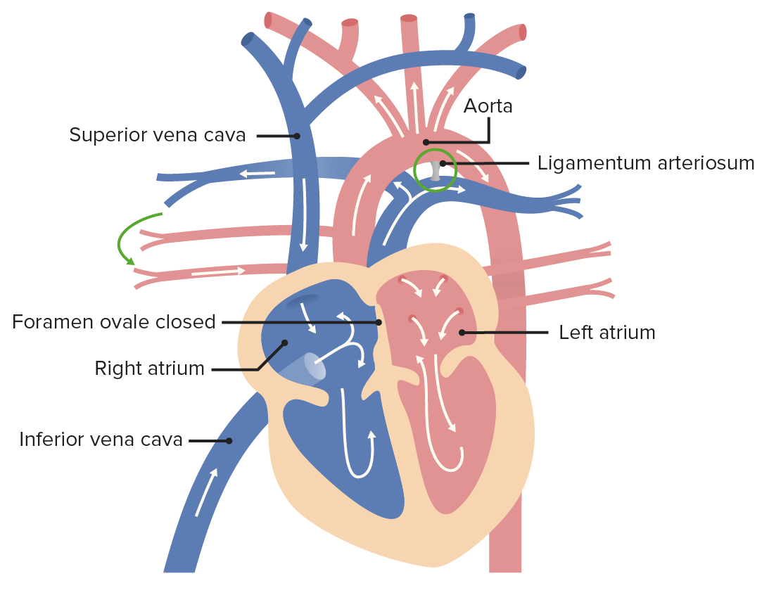
00:01 Hello, and this is the first talk on how the heart sub-divides to form the four-chambered heart and we'd get a single atrium transitioning to become the right and left atria. 00:12 Now, there are gonna be several linked talks with this one. 00:15 Splitting of the ventricles, splitting of the atria from the ventricles, and partitioning of the outflow tracks. 00:22 We're gonna address each of this topics separately but it's important to underscore all of these processes are happening at the same time. 00:29 So we're gonna start with partitioning of the atrium, On the left image, we are looking at the view of the heart from the right. So we are looking from the right atrium towards the left part of the atrium. And on the right side of the screen, we are looking at the heart from its anterior view. So we’ve taken the anterior surface of the heart off in the coronal plane and were looking at it from the front. 00:53 And on the right side of the screen, we're looking at the heart from its anterior view. 00:59 So we've taken the anterior surface of the heart off in the coronal plane and were looking at it from the front. 01:05 Now, as development moves along, the sinus venosus migrates its opening towards the right side of the atria. 01:12 So instead of equally emptying into the right and left sides of the atrium, it transitions to the right. 01:18 And that's gonna be important because the flow of blood, thereafter, has to move from the right side of the atrium to the left side of the atrium, as is indicated by the red arrow. 01:27 Now, early on, the atria develops a little down growth, a little wall that grows down called the septum primum, meaning the first wall. 01:37 So the septum primum is gonna be the first thing that starts to divide the atrium into a right and a left atrium. 01:43 The hole that's left below that is called, the ostium primum or first hole. 01:48 So that's gonna be what starts, it's going to meet a little division that's developing between the atrium and the ventricles. 01:55 There's gonna be a dorsal and ventral endocardial cushion present there. 02:00 Now on the right, we're showing the cut with that dorsal and endocardial cushion in the plain of the cut. 02:05 But it's important to know that the right and left atria are both equally open to the right and left ventricles, at least initially. 02:12 So as development proceeds, that septum primum appears to grow down, down, down, and the ostium primum gets smaller and smaller. 02:22 Now, as the dorsal and ventral endocardial cushions grow closer together, they're gonna narrow that ostium primum tremendously. 02:31 And that's gonna cause a major problem, because if the blood cannot flow from the right side of the atrium to the left side, we've interrupted blood flow through the embryonic heart, broken our cardinal rule and the embryo is going to die. 02:44 So how does the body fix this? It fixes this by making another hole. 02:50 The ostium primum gets narrower and narrower, but small little holes develop in the septum primum, that are going to enlarge and eventually perforate to become a single hole called the ostium secundum, the second hole. 03:02 So as the ostium primum gets narrower and narrower, a little more cranially, we get the ostium secundum. 03:09 And that allows blood flow to continue from right to left across the atria. 03:13 So the ostium primum getting smaller and smaller, ostium secundum getting bigger and bigger. 03:20 And one thing to note, at this point is, we've got oxygenated blood coming from the placenta up the inferior vena cava into the right atrium and from there it passes into the left part of the atrium. 03:33 And that well-oxygenated blood then passes down the left side into the developing ventricle. 03:40 Now nearby, we've got the superior vena cava bringing blood into the right atrium as well. 03:47 And that poorly oxygenated blood is preferentially directed down to the right side of the ventricle which is going to be at least at first, the bulbus cordis. 03:58 So we've already got a little bit of a partitioning of oxygenated blood to the left and poorly oxygenated blood to the right. 04:04 So the ostium primum gets smaller and smaller as the endocardial cushions grow together, and we start to get an atrioventricular septum. 04:14 And eventually, we've got nothing but the ostium secundum in the septum primum. 04:19 Now, all seems well enough but we get something else happening. 04:24 That ostium secundum starts to get closed over by another structure, that's gonna be called the septum secundum. 04:35 The septum secundum or the second wall is going to grow in and start to cover the ostium secundum. 04:40 And a little preview, that's going to risk shutting of blood flow through the embryonic heart. 04:45 So let's see what the embryo does to deal with that. 04:48 What it's going to do is keep getting larger. 04:54 And the septum secundum comes from the fact, that to get larger, the atria literally have to pull parts of the sinus venosus into their wall. 05:02 So as the right atrium gets larger, it pulls the sinus venosus and part of the venous system into its wall, that's shown here in purple, and it's gonna move the superior and inferior vena cava closer and closer into the right atrium until they have their own separate opening. 05:19 So, initially, the sinus venosus has a single opening into the right side and the right atrium receives that blood. 05:27 Better oxygenated blood coming from the inferior vena cava and placenta, poorly oxygenated blood only coming from the superior vena cava. 05:34 Now note, that on left side, we have an opening for the pulmonary vein, the blood flow from the lungs. 05:41 As the left side needs to get larger it's gonna pull the wall that pulmonary vein in until it actually pulls the whole wall in and will have four separate pulmonary veins as tributaries. 05:53 So little further along, purple is showing where the sinus venosus got pulled into the right atrium and lighter purple is gonna be showing where pulmonary vein got pulled into the left atrium. 06:05 Now, in the process of moving into the right atrium, the sinus venosus forms a wall and that is that same septum secundum we saw just a bit earlier. 06:17 So let's return to a little bit more similar picture to what we saw before but one thing before we move on I wanna note is that as the sinus venosus moves in, we got separate openings for the superior vena cava, inferior vena cava, and the blood drainage from the heart itself, the coronary sinus. 06:36 And that's all going into the right atrium. 06:38 And pulmonary veins to the left atrium. 06:41 That's gonna be more or less how the adult atria are laid out. 06:45 So let's return here. 06:47 The septum secundum has enlarged and it's now covering the foramen secundum. 06:56 That's a problem because we're gonna start to narrow that flow and prevent blood from flowing from right to left and kill the embryo. 07:04 The way that we're gonna deal with this is because it doesn't quite cover the septum primum. 07:12 There's a tiny little flap hanging out there. 07:15 Now the remaining hole in the second wall, the septum secundum, is called, the foramen ovale or oval foramen. 07:23 So the oval foramen is a gap in the septum secundum. 07:27 That means we can see the septum primum from there. 07:31 And as blood flow enters the right atrium, It's going to push that portion of the septum primum out of the way and allow blood to continue traveling from right to left. 07:45 So even though it looks like, in the picture on the right, that the septum secundum has completely blocked the atrium, there is still some blood flow that's pinching its way through that little narrowed section of the foramen ovale. 07:58 So how does development allow this to happen and have blood flow move to the atria? The good news is that foramen ovale is fairly broad and the septum primum that's up against it is very membranous, it's very flimsy. 08:14 And so as blood entering the inferior vena cava comes into the right atrium it's going to be directed to hit the septum primum and it's gonna push that portion of the septum primum out of the way. 08:28 And that's gonna be now known as the valve of the foramen ovale. 08:32 Meaning, the foramen ovale is the hole in the septum secundum, the blood hits that valve, the valve opens and well-oxygenated blood from the placenta and inferior vena cava is preferentially directed to the left atrium and thereafter, the left ventricle. 08:48 Poorly oxygenated blood coming from the superior vena cava goes into the right atrium but it has a straight shut down towards what's gonna become the right ventricle of the heart. 08:59 So this is what allows blood to flow through the embryonic heart, but when we're born, the pressure inside the left atrium is gonna increase as blood flow to the pulmonary circuit increases, more blood coming to the pulmonary veins into the left atrium will slap that valve of that foramen ovale shut and that's what's gonna functionally separate our right and left atria. 09:22 So let's just take a second. 09:24 Pass all that crazy terminology to appreciate the fact that our body has come up with a hack to get around the fact that we need to have a linear flow of blood throughout our entire embryonic development, yet transition to a separate systemic and pulmonary circuit the minute we take our first breath. 09:41 Pretty amazing.
About the Lecture
The lecture Formation of the Right and Left Atria by Peter Ward, PhD is from the course Development of Thoracic Region and Vasculature. It contains the following chapters:
- Formation of the Right and Left Atria
- Expansion of the Atria and Development of the Foramen Ovale
Included Quiz Questions
What structure descends from the roof of the atrium and partially separates the left and right atrium?
- Septum primum
- Septum secundum
- Ostium primum
- Septum transversum
- Endocardial cushion
The endocardial cushions are derived from what embryonic cell layer?
- Neural crest
- Lateral plate mesoderm
- Intermediate mesoderm
- Cardiogenic mesoderm
- Endoderm
Apoptosis of the septum primum yields what structure?
- Ostium secundum
- Septum secundum
- Ostium primum
- Septum transversum
- Fossa ovalis
The upper portion of the septum secundum arises from what heart structure?
- Sinus venosus
- Left atrium
- Right atrium
- Endocardial cushion
- Bulbus cordis
What structure found in the septum secundum allows blood to flow from the right atrium to the left atrium?
- Foramen ovale
- Septum primum
- Ostium primum
- Septum secundum
- Fossa ovalis
Author of lecture Formation of the Right and Left Atria

Peter Ward, PhD
Customer reviews
4,0 of 5 stars
| 5 Stars |
|
1 |
| 4 Stars |
|
1 |
| 3 Stars |
|
1 |
| 2 Stars |
|
0 |
| 1 Star |
|
0 |
First time watching it and it's really well explained. Some things are hard to understand, but it helps to pause and try to imagine it. Couldn't ask for more. Educating videos shouldn't be movies, pausing to understand works fantastic as everything explained makes sense.
it is really good and easy to understand i watched it twice and i memorized everything but in the quiz there was a question that went like what are the endocardial cushions derived from and in the lecture there was no mentions of such thing
Nice lecture, but i would understand this better if there were 3D animation videos....That would make it more visual and interesting...
