Playlist
Show Playlist
Hide Playlist
Boundaries and Muscles of the Posterior Abdominal Wall

-
Slides Boundaries and Muscles of the Posterior Abdominal Wall.pdf
-
Download Lecture Overview
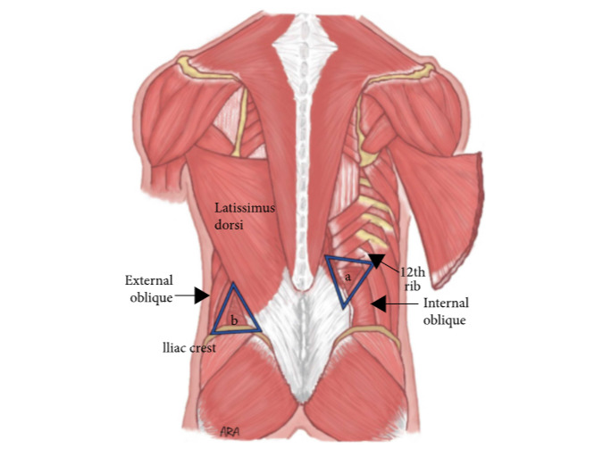
00:01 So this topic is going to be about the posterior abdominal wall. 00:06 So let's start by looking at the boundaries of the posterior abdominal wall, and then talk about the diaphragm. 00:12 So here you'll be familiar with this image from previous slides. 00:16 And you can see that the posterior abdominal wall is a highly muscularised structure. 00:22 There's lots of muscles that are running down either side of the vertebral column. 00:27 And these form an important support network to help maintain our posture. 00:33 Let's just have a look at the boundaries of the posterior abdominal wall. 00:36 And here most posteriorly, we've got the lumbar vertebrae, one through to five indicated. 00:42 You can also see we have the 12th rib highlighted here in green, and also the iliac crest. 00:48 And these helped to form part of the boundaries of that posterior abdominal wall. 00:53 There's some other important landmarks like this part of the iliac fossa. 00:58 And we can also see it extends inferiorly to the pelvic brim. 01:02 We'll talk about that later on when we look at the pelvis. 01:05 But now we can bring all of that together and see an outline of the posterior abdominal wall. 01:12 Posteriorly, now we can clearly make out the boundaries. 01:16 We can see the green highlighted vertebral column, the posterior aspect of the 12th rib, and also the iliac crest. 01:22 And here these lines indicate the boundaries of that posterior abdominal wall from the posterior aspect. 01:29 Here we can see sitting most superiorly and extending up into the thoracic cage, and really forming the upper limit of the abdomen. 01:38 We have the diaphragm. 01:39 And again indicates we've got the 12th rib, which is an important attachment site for the diaphragm. 01:45 These together form the superior border of the posterior abdominal wall. 01:50 Remembering that the five lumbar vertebrae from the posterior abdominal wall, his posterior border. 01:57 And we can see that inferiorly, we have that pelvic brim that we mentioned a moment ago. 02:02 So those structures we spoke about these helped to form various boundaries of the posterior abdominal wall. 02:08 Now we can add on the musculature that occupies this space. 02:13 We have some important muscles. 02:15 We have psoas major here and running anterior to psoas major, we have psoas minor. 02:21 Sitting lateral to psoas minor, we have quadratus lumborum, and these are important muscles. 02:27 As we extend down into the pelvis, we pick up iliacus muscle that lines the inside of the pelvic wall. 02:35 So now let's start looking at these muscles individually. 02:38 Here on the screen we can see we have psoas major that sits lateral to the lumbar vertebrae, and also the 12th thoracic vertebrae as well. 02:47 We can see that it originates here from the transverse processes of the lumbar vertebrae. 02:52 And here we can see the bodies of the lumbar vertebrae and also the 12 thoracic vertebrae alongside the intervertebral discs. 03:00 It originates from this location and passes posteriorly and inferiorly all the way down to the lesser trochanter of the femur. 03:09 Let's have a look at the nerve supply to the psoas major muscle. 03:13 And this is coming from the anterior rami of the spinal nerves L1 through to L3. 03:19 And these go on to supply the psoas major muscle. 03:23 The muscle is important in helping to flex the trunk when you're in a sitting position. 03:27 So to bend your trunk forwards and also it helps to flex the thigh. 03:31 So to bring the thigh forwards as well. 03:35 Sitting directly anterior to psaos major is psoas minor and as its name suggests, this is a much smaller muscle. 03:43 And it may not always be present in some people, but it is quite common. 03:48 Psoas minor, as I said, is a smaller muscle that sits directly anterior to psoas major. 03:54 It originates only from the bodies of T12 and L1, and here we can see the intervertebral disc between those two vertebrae. 04:02 It runs all the way down to innovate, to insert into the pectineall line of the pelvis. 04:08 The nerve supply to psoas minor is similar to that of psoas major, but it's really only the first rami of L1, the anterior rami of L1 the first lumbar vertebrae, and that provides the innovation to psoas minor. 04:23 Psoas minor which is like I said consistent but also regularly seen also helps to flex the vertebral column but it's quite a weak flexor as its size would lead to. 04:34 We can see here if we move now down into the pelvis, we're looking at iliacus muscle. 04:39 And this very much sits on the lateral aspects of the pelvic wall. 04:44 We can see it's coming from the iliac crest and it passes all the way down to the lesser trochanter of the femur. 04:52 The innovation of the iliacus muscle is by way of the femoral nerve. 04:57 Iliacus muscle is important as it helps to flex the thigh. 05:03 Together the iliacus muscle and the psoas major muscles combine to have a joint tendon. 05:09 And here we have what's called iliopsoas, where these two muscles pass down and they run towards the lesser trochanter of the femur. 05:18 The innovation of the iliopsoas muscle is the same for both psoas major and the iliacus muscle individually, but their tendons importantly combined. 05:30 Now, let's talk about quadratus lumborum. 05:32 Quadratus lumborum originates from the iliac crest on the ilium. 05:37 And it passes superiorly and medially to insert into the transverse processes of the lumbar vertebrae, and also the inferior surface of the 12th rib. 05:48 Quadratus lumborum is an important muscle and is innovated by the anterior rami of T12. 05:55 And L1 through to L4, as you can see here. 06:00 Quadratus lumborum has an important job working independently as they help to laterally flex the vertebral column. 06:07 So if one quadratus lumborum muscle works independently, it works on its own, then it will help to laterally flex the vertebral column. 06:16 If however, both of these muscles were to work together, then it actually helped to extend the vertebral column. 06:22 So they antagonize the work of psoas major muscle.
About the Lecture
The lecture Boundaries and Muscles of the Posterior Abdominal Wall by James Pickering, PhD is from the course Posterior Abdominal Wall.
Included Quiz Questions
Which muscle is NOT part of the posterior abdominal wall?
- Transversus abdominis
- Quadratus lumborum
- Psoas major
- Iliacus
- Psoas minor
Which action does the quadratus lumborum perform?
- Lateral flexion of the vertebral column
- Extension of the thigh
- Flexion of the thigh
- Medial rotation of the vertebral column
- Medial rotation of the thigh
A patient had a severe accident in which his pelvis was fractured. Now he complains of an inability to flex his thigh. Which nerve roots are most likely damaged?
- L2, L3, and L4
- L1 and L2
- L2 and L3
- L4 and L5
- L3 and L4
Which structure passes through the esophageal hiatus?
- Vagus nerve
- Left phrenic nerve
- Internal thoracic artery
- Greater splanchnic nerve
- Thoracic duct
Author of lecture Boundaries and Muscles of the Posterior Abdominal Wall

James Pickering, PhD
Customer reviews
5,0 of 5 stars
| 5 Stars |
|
5 |
| 4 Stars |
|
0 |
| 3 Stars |
|
0 |
| 2 Stars |
|
0 |
| 1 Star |
|
0 |
