Playlist
Show Playlist
Hide Playlist
Axillary Artery

-
Slide Axillary Artery.pdf
-
Download Lecture Overview
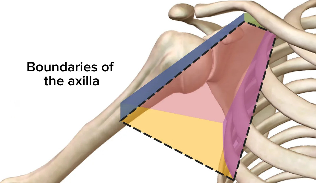
00:01 So, let's start by having a look at the axillary artery. 00:05 So, the axillary artery really is the direct continuation of the subclavian artery. 00:10 Here, you could see the subclavian artery passing out of the brachiocephalic trunk from the heart. 00:15 And in this case, it's coursing towards the right-hand side where it enters into the axilla. 00:22 Here, we can see the lateral margin of the first rib is the landmark where the subclavian then becomes the axillary artery. 00:29 The axillary artery then, runs through the axilla and at the lower border of teres major, it becomes the brachial artery. 00:37 Using the pectoralis minor muscle as an important landmark, this can separate the axillary artery into three parts. 00:44 The first part is situated between the superior boundary of pectoralis minor and the clavicle where it's coming from the subclavian artery. 00:53 Here, we can see the second part which is deep to the pectoralis minor muscle. 00:57 The third part of the axillary artery is really appearing between the inferior border of pectoralis minor muscle and the inferior border of teres major muscle. 01:08 And this is where we find the third part of the axillary artery. 01:12 There are a number of arteries that come off these respective parts. 01:15 So, coming from the first part of the axillary artery, we have the superior thoracic artery. 01:20 Coming from the second part of the axillary artery, we have the thoraco-acromial artery. 01:26 We also have the lateral thoracic artery that passes down the lateral aspects of the thoracic cage. 01:32 And then, moving to the third part of the axillary artery, we have the subscapular artery. 01:37 And then, importantly, we have both the anterior and posterior circumflex humeral arteries. 01:44 And these go around the surgical neck of the humerus. Dislocation of the humeral head, so, the head of the humerus if it becomes dislocated can actually start compressing onto the axillary artery. 01:57 So, it can jam the axillary artery between the dislodged humeral head against the first rib and the lateral wall of the rib cage. 02:06 And this can lead to occlusion of that blood vessel, preventing blood from passing distally towards the upper limb. 02:13 If we then look at the thoraco-acromial arteries specifically, we have an acromial branch that goes to supply regions around the acromion and the joint, the clavicular acromial joint for example. 02:23 And we also have a clavicular branch that goes on to supply the clavicle. 02:27 We have a pectoral branch that supplies the pectoralis muscles, so, pectoralis minor, pectoralis major. 02:33 And we also have another branch supplying the mass muscle which is the deltoid muscle, forming the real shape of the shoulder that we can see in the living person. 02:43 So, we have a deltoid branch coming off the thoraco-acromial artery. 02:48 This is a really important artery that supplies lots of things around the shoulder joint. 02:52 It also gives rise to that lateral thoracic artery which we can see passing down on the lateral aspect of the thoracic cage. Here, we can see it passing down here. 03:02 So, we have a lateral thoracic artery which we can see here, supplying serratus anterior muscle. 03:08 We can also find the subscapular muscle. 03:11 So, the subscapular muscle is going to work its way posteriorly to supply subscapularis muscle. 03:16 And here, we can see it doing so as it courses towards the scapular, supplying subscapularis, the muscle on the anterior surface of the scapula. 03:26 We can see it gives rise to the thoracodorsal artery which we can see here. 03:30 And we have various other named branches, the circumflex scapular artery. 03:34 And that's passing through the triangular space as that space that allows structures to leave the axilla and pass through the posterior aspect of the shoulder region. 03:44 Here, we could now see the circumflex scapular artery passing up along the posterior surface of the scapula humeral muscles. We can see it running up infraspinatus. 03:55 The dorsal branch of the dorsal scapular artery is an important branch that comes down to join that circumflex scapular artery and here, we have an important anastomosis. 04:05 Helping when we spoke previously about axillary artery being occluded, perhaps, due to dislocation. 04:11 It means that use these anastomosis, blood can still reach this region by an alternative root. 04:16 So, these anastomosis can be important. 04:19 Another important anastomosis that we need to be aware of is around the circumflex humeral arteries. 04:24 Both the anterior and posterior branches run around the surgical neck where they form this important anastomosis. 04:32 So, here, you can see the surgical neck of the humerus and the posterior and anterior circumflex humeral arteries are converging to form that anastomosis around this region. 04:43 Remember that the posterior circumflex humeral artery leaves the axilla by passing through the quadrangular space.
About the Lecture
The lecture Axillary Artery by James Pickering, PhD is from the course Fasciae and Neurovasculature of the Upper Limbs.
Included Quiz Questions
Which artery branches from the first part of the axillary artery?
- Superior thoracic artery
- Lateral thoracic artery
- Thoracoacromial artery
- Anterior circumflex humeral artery
- Posterior circumflex humeral artery
What injury may lead to axillary artery occlusion?
- Humeral head dislocation
- Radius fracture
- Scapula dislocation
- 4th rib fracture
- 7th rib fracture
What best describes the location of the second part of the axillary artery?
- Deep to pectoralis minor muscle
- Superficial to pectoralis minor muscle
- Deep to pectoralis major muscle
- Superficial to pectoralis major muscle
- Lateral to pectoralis major muscle
How many branches leave the thoracoacromial artery?
- 4
- 3
- 2
- 1
- 5
Author of lecture Axillary Artery

James Pickering, PhD
Customer reviews
5,0 of 5 stars
| 5 Stars |
|
5 |
| 4 Stars |
|
0 |
| 3 Stars |
|
0 |
| 2 Stars |
|
0 |
| 1 Star |
|
0 |
