Playlist
Show Playlist
Hide Playlist
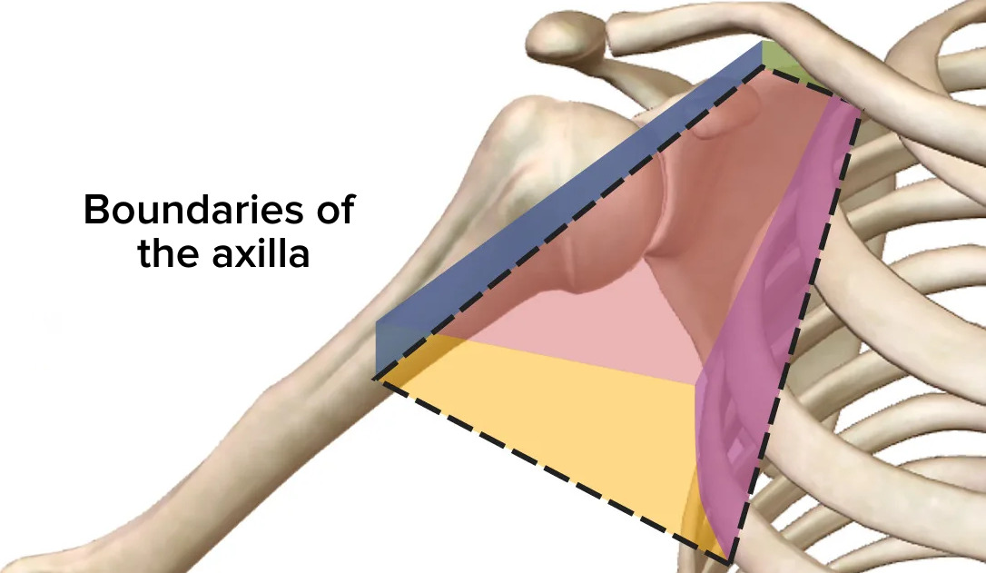
Walls and Boundaries of the Axilla

-
Slide Axilla Walls and Boundaries.pdf
-
Download Lecture Overview
00:01 In this lecture, we're going to look at the axilla and brachial plexus. 00:05 So, let's start by looking at the axilla, an important space in the superior aspect of the upper limb. 00:14 So, before we look at the axilla, let's just remind ourselves of some important bony structures around this region. 00:21 So, highlighted on the screen in green, we have the humerus. 00:25 We then, have the scapula more posteriorly. Here, we have the clavicle at the superior aspect. 00:31 And then, we have some of the ribs, ribs one through to five, you could see highlighted on the lateral aspect of this upper part of the thoracic wall. 00:41 And the axilla really is a space. It's a fat-filled space that contains a number of important blood vessels and nerves and lymphatic structures which we'll look at in a few moments time, look past between the neck and the upper limb. 00:58 It's a space therefore that has a number of boundaries. It has an apex and a base. 01:03 It then, has four walls which are the medial wall, the lateral wall, the posterior, and the anterior wall. 01:12 And these boundaries form that axilla, this important space at the superior aspect of the upper limb. 01:21 So, let's just remind ourselves of this structure in a little bit more detail with some muscles now included. 01:27 So, here, we can see looking down onto the superior aspects, we can see we have the apex of the axilla highlighted here and we could see, we have the boundaries being the first rib medially. And then, anteriorly, we have the clavicle. 01:43 And then, here, we have the superior border of the scapula on the posterior aspect. 01:49 It contains a number of important structures that are passing from the neck into this axilla. 01:54 Highlighted in blue, we have the subclavian vein. Then, we have the subclavian artery. 01:59 And we have an incredibly important network of nerves. This is called the brachial plexus. 02:06 So, these structures are passing from the neck region into the upper limb or as the venous structure indicates, passing from the upper limb into the neck region. 02:16 And they do this by passing through that space which is the axilla. 02:20 They run very closely as you can see on the diagram here to the first rib. 02:24 And therefore, any damage to the first rib, perhaps, by a fracture or something, can actually damage some of these vessels. 02:31 Importantly, it can damage the subclavian artery which runs very close to this structure. 02:38 Here, we can see some of the bony makeup now of the axilla. So, anteriorly, we have pectoralis major. 02:44 We also have that small muscle underneath the clavicle, subclavius and we also have pectoralis minor. 02:51 These muscles passing from the chest wall, across to the upper limb, forming the anterior boundary. 02:57 We also have that sheet of fascia, the clavipectoral fascia that is running all the way across the anterior aspect of the axilla. 03:06 Medially, we have a couple of bone - a couple of muscular structures. 03:09 We have the various muscles that make up the thoracic wall, including the ribs. 03:15 So, we have the intercostal muscles and we also have serratus anterior, that important muscle on the lateral aspects of the chest wall. 03:24 Laterally, we have the intertubercular sulcus of the humerus. 03:28 We'll come back to this when we look at the muscles in the arm as that's an important pathway for various tendons and that forms part of the lateral wall. 03:37 Posteriorly, we then have an important muscle which is subscapularis that lies on the anterior surface of the scapula. So, here, we have subscapularis muscle. Also on the posterior wall, we have triceps brachii and we have teres major muscles. 03:52 Some important muscular structures that form the posterior wall. 03:56 Most inferiorly of this group, we have latissimus dorsi as well. 04:01 The floor of the axilla, if we could now see with the anterior wall put back in place, the floor of it really is the skin of the armpit region. 04:10 And you can grab this aspect if you were to pinch the skin flap that's connecting your chest wall to your arm. 04:16 And that's really the armpit region. And this forms the floor of the axilla. 04:21 Passing through the axilla as I've mentioned before, there's a whole series of structures. 04:28 And we can see now, within that space that's being formed by these various landmarks, we have the brachial plexus, the subclavian artery and the subclavian vein, and some important lymphatic structures.
About the Lecture
The lecture Walls and Boundaries of the Axilla by James Pickering, PhD is from the course Anatomy of the Axilla.
Included Quiz Questions
Which statements about the boundaries of the axilla are correct? Select all that apply.
- The apex is formed by the clavicle, 1st rib, and superior border of the scapula.
- The medial wall is formed by the pectoralis major muscle.
- The anterior boundary is formed by the pectoralis major and minor.
- The posterior wall is formed by the subscapularis muscle, the scapula bone, teres major, and latissimus dorsi muscle.
- The lateral wall is formed by the intertubercular groove of the humerus.
How many important spaces are present within the axilla along the posterior wall?
- 3
- 2
- 4
- 5
- 6
Which structures are included in the contents of the axilla? Select all that apply.
- Axillary artery
- Pectoralis major and minor muscles
- Axillary vein
- Brachial plexus
- Axillary lymph nodes
Author of lecture Walls and Boundaries of the Axilla

James Pickering, PhD
Customer reviews
5,0 of 5 stars
| 5 Stars |
|
5 |
| 4 Stars |
|
0 |
| 3 Stars |
|
0 |
| 2 Stars |
|
0 |
| 1 Star |
|
0 |
