Playlist
Show Playlist
Hide Playlist
Skull: Introduction

-
Slides Anatomy Skull Introduction.pdf
-
Download Lecture Overview
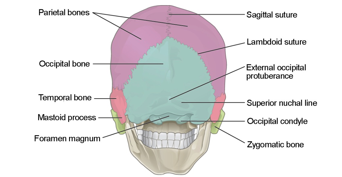
00:01 Let's start a discussion of head anatomy by focusing on the bones of the skull. 00:08 So here we have a lateral view and a superior view. 00:13 And together, the bones of the skull can be divided into a neurocranium, because they're the bones that essentially house the brain and then a visceral cranium, which are more like the bones of the face. 00:29 In terms of individual bones, the first bone we see here anteriorly is the frontal bone. 00:36 Then we see a bone in the area of the nose called the nasal bone that will be attaching to cartilage, more anteriorly. 00:43 We can see a little bit of the lacrimal bone here and lacrimal refers to tears. 00:48 So this is a bone that's going to have a duct for the passageway of tear fluid. 00:53 We can see the outer portion of the sphenoid bone which has a very complicated shape internally. 01:00 We see the maxilla and the zygomatic bone in the area of the cheeks. 01:07 Then we have the jaw bone, which we call the mandible. 01:11 Very complicated looking bone called the temporal bone. 01:15 And then most posterior in inferiorly, we have the occipital bone and forming a large part of either side of the skull, or the parietal bones. 01:28 Now, there are some features that we're going to point out such as the zygomatic arch, which isn't actually a distinct bone. 01:36 It's actually an arch formed from parts of two different bones, both the zygomatic bone and the temporal bone. 01:44 We also see this pointy projection here coming off of the temporal bone that we call the styloid process, a more rounded projection off the temporal bone that we call the mastoid process. 01:57 And then also an opening in the temporal bone called the external acoustic meatus. 02:02 And as the name implies, this is going to be the passageway for sound to travel. 02:08 There's also a bump or projection on the occipital bone. 02:13 That's a landmark known as the Inion. 02:17 If we swing around to an anterior view, we see the frontal bone most prominently. 02:23 We also see the nasal bones pretty well where they would attach to the nasal cartilage, a little bit of the lacrimal bone, and pretty much all of the maxilla here. 02:34 We also see a large portion of the mandible or the jaw, a little bit of the zygomatic bone, a little bit of the sphenoid bone, and only a tiny amount of the palatine bone which can be very deep inside the skull. 02:48 We also see another very deep bone called the ethmoid bone through the opening of the nasal cavity. 02:55 We don't see much of the temporal bone because it's off to the side. 02:59 Nor do we see much of the primal bone because it's also very laterally oriented. 03:06 The bones of the skull are largely formed by non movable joints that we call sutures. 03:13 The first suture here between the frontal and parietal bones lies in the coronal plane, hence we call it the coronal suture. 03:22 We also have the suture between the temporal bone in the parietal bones called the squamous suture. 03:28 And we call it that because the flat part of the temporal bone is called the squamous portion. 03:35 We also have a suture between the occipital bone and parietal bones that vaguely resembles the Greek lambda character. 03:43 So it's called the lambdoid suture. 03:46 And then in the midline, running in the sagittal plane between the left and right parietal bones is the sagittal suture. 03:54 We also have landmarks such as the bregma, between the coronal and sagittal sutures, and the lambda between the lambdoid and sagittal sutures. 04:05 And these landmarks during development, were actually fairly soft areas and they were the soft spots on an infant skull known as the anterior and posterior fontanelles. 04:16 Speaking of development, there was also a suture between the left and right parts of the frontal bone called the frontal or metopic suture. 04:23 It's usually the first one to fuse and often disappears, but sometimes remnants of that metopic suture can be seen in the frontal bone. 04:34 There are some smaller sutures that are more descriptive in their names, such as the suture between the frontal and sphenoid bone called the sphenofrontal. 04:43 We also have a small one between sphenoid and parietal called sphenoparietal. 04:48 And then a small one between the squeamish portion of the temporal bone and the sphenoid called the sphenosquamous. 04:55 Further back in the area of the mastoid, we have the parietal mastoid and occipitomastoid. 05:03 Where all of these small sutures meet in the sphenoid area is also a landmark called the pterion and a similar one in the mastoid area is called the asterion.
About the Lecture
The lecture Skull: Introduction by Darren Salmi, MD, MS is from the course Skull.
Included Quiz Questions
Which of the following bones is the most anterior?
- Maxilla
- Temporal
- Occipital
- Sphenoid
- Parietal
Which suture lies between the frontal and parietal bones of the skull?
- Coronal
- Squamous
- Lamboid
- Lambda
- Transverse
Which of the following bones is the most inferior?
- Mandible
- Frontal
- Sphenoid
- Nasal
- Lacrimal
Author of lecture Skull: Introduction

Darren Salmi, MD, MS
Customer reviews
5,0 of 5 stars
| 5 Stars |
|
5 |
| 4 Stars |
|
0 |
| 3 Stars |
|
0 |
| 2 Stars |
|
0 |
| 1 Star |
|
0 |
