Playlist
Show Playlist
Hide Playlist
Posterior Compartment of the Thigh


-
Slide Posterior Compartment of the Thigh.pdf
-
Download Lecture Overview
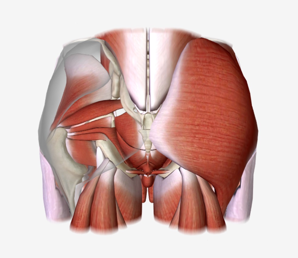
00:01 Now let's have a look at these muscles in a bit more detail. 00:03 So let's have a look at the posterior thigh muscles. 00:07 Here we can see a number of muscles that form in this location. 00:10 We have semimembranosus, and we have a similarly named muscle known as semitendinosis. 00:16 The difference between these two apart from their positioning is that the semitendinosis has a very thick tendon that actually starts quite more proximately than semimembranosus, which has a slightly flatter muscle helpings, give them their two names. 00:30 So running on the medial aspect, we have semimembranosus, semi tendinosis, and then running more on the lateral aspect, we have biceps femoris. 00:39 As its name implies, like biceps brachii, it has two heads. 00:43 Let's have a look at semibranosus, the origin and insertion of semimembranosus. 00:49 The origin is it comes down from the ischial tuberosity and it goes and attaches to the posterior surface of the medial tibial condyle. 00:58 So we see it runs distally down towards the leg. 01:02 Semi tendinosis, this also comes from the ischial tuberosity. 01:05 It lies more superficial than semimembranosus. 01:08 But it also goes and attaches to the tibia. 01:11 This time is attaching to the medial surface of the proximal tibia. 01:16 So it's attaching just medial to the tibial tuberosity. 01:21 Biceps femoris is the more laterally positioned muscle in the posterior thigh. 01:25 And here we can see it's long head, remember biceps means it has two head, its long head is coming from the ischial tuberosity. 01:33 It also has a shorter head and this is coming from the linear aspera. 01:37 Remember that line on the posterior aspect of the femur. 01:41 So the two heads, long head, short head, coming from the ischial of tuberosity and the linea aspera respectively. 01:48 And then passes down and attaches to the head of the fibular. 01:53 These muscles are supplied by the sciatic nerve, but primarily they also have an important function in movement of the knee. 02:02 So here we can see the position of the hamstring muscles is sitting very much posterior to the femur. 02:09 Now what this means is, as it runs posterior to the femur, it runs posterior to the knee joint. 02:15 And as it actually runs posterior to the knee joint, it is going to flex the knee as it moves the knee backwards. 02:22 So here we see flexion of the knee joint as the hamstring muscles cross the knee joint and attach to the neck. 02:28 So that's going to flex the knee. 02:30 As they also cross the hip joint as they're attaching to the ischial tuberosity, contraction of these muscles will also lead to extension of the hip joint. 02:40 So the hamstrings are important in flexing the knee and also extending the hip joint. 02:46 If we have a look at the neurovasculature of the posterior thigh, here we can remove biceps to see clearly sciatic nerve and we can see a number of various perforating arteries that are coming from the deep femoral artery and these are running down and how many to supply the deep substance of these muscles.
About the Lecture
The lecture Posterior Compartment of the Thigh by James Pickering, PhD is from the course Anatomy of the Thigh.
Included Quiz Questions
What is the insertion site for the semimembranosis?
- Posterior surface of the medial tibial condyle
- Anterior surface of the medial tibial condyle
- Anterior surface of the lateral tibial condyle
- Posterior surface of the lateral tibial condyle
- Ischial tuberosity
What is the origin of the long head of the biceps femoris?
- Ischial tuberosity
- Linea aspera
- Head of fibula
- Posterior surface of the lateral tibial condyle
- Anterior surface of the lateral tibial condyle
What nerve innervates the biceps femoris?
- Sciatic
- Femoral
- Tibia
- FIbula
- Axillary
Author of lecture Posterior Compartment of the Thigh

James Pickering, PhD
Customer reviews
5,0 of 5 stars
| 5 Stars |
|
5 |
| 4 Stars |
|
0 |
| 3 Stars |
|
0 |
| 2 Stars |
|
0 |
| 1 Star |
|
0 |
