Playlist
Show Playlist
Hide Playlist
Forearm in Cross Section

-
Slide Forearm in Cross Section.pdf
-
Download Lecture Overview
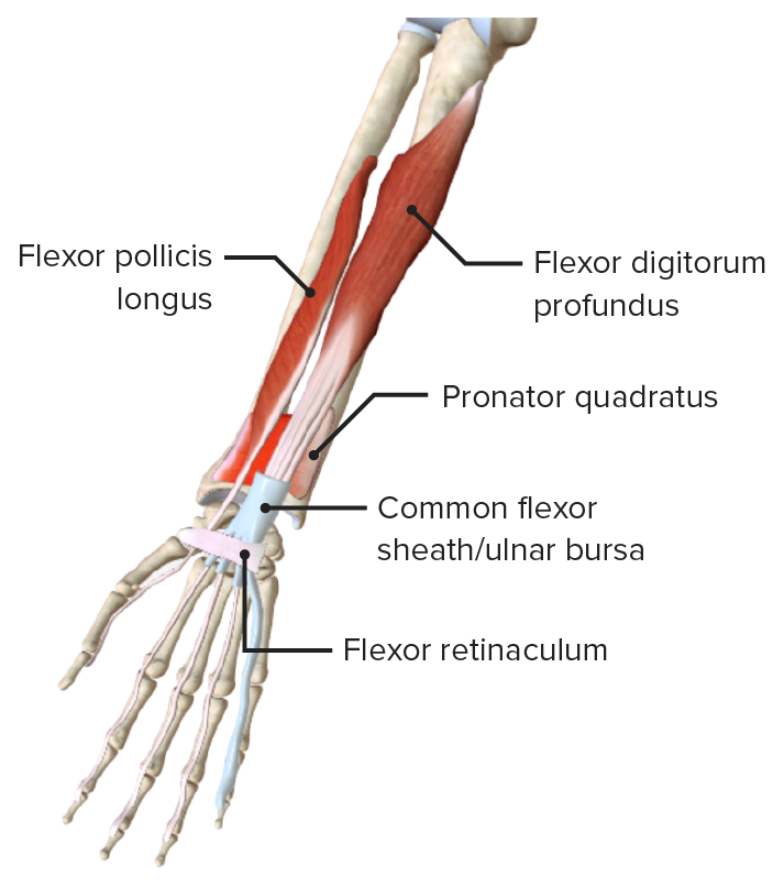
00:01 In this lecture, we'll look at the more distal area of the upper limb, concentrating on the forearm. 00:08 So, let's start off by looking at the forearm in cross-section, just like we did when we looked at the arm. 00:14 So, similarly, we have a cross-section through the forearm which is as if the patient is supine, lying on their back, and you're looking at them from the feet end of the person. 00:26 So, when you're looking up through the arm at this cross-section. 00:30 Importantly, we can see running through the forearm, we have two bones. 00:33 We have the ulna and we have the radius, connected via the interosseus membrane we can see highlighted there. 00:40 And similarly to the brachial fascia, we have the antebrachial fascia running all the way around the forearm, separating this muscular compartment from the subcutaneous tissue. 00:52 This similarly to the arm also has a number of penetrating layers. 00:57 So, here, we could see the lateral intermuscular septum. 01:00 And here, we can see the medial intermuscular septum, running towards the radius and the ulnar bones respectively. 01:07 And what this does is this helps to create the anterior compartment and the posterior compartment. 01:13 And in the anterior compartment, we can see we have a number of muscles. 01:16 In this posterior compartment, we see we have a number of muscles also. 01:21 So, now, let's have a look at the various muscles that fall into these compartments. 01:26 We're not going to go into detail, it's just to start familiarizing you with the various words that make up these muscles. 01:32 Because there's a lot here. So, just take your time and go through them slowly. 01:36 In the anterior compartment, we have flexor digitorum profundus, flexor carpi ulnaris, flexor digitorum superficialis, and flexor pollicis longus. 01:48 As the name implies, these are muscles which are flexor. 01:52 So, they help to flex various joints occurring distally within the forearm, even passing into the hand. 01:59 So, flexor is an important word there. We then have words like carpi for the carpal bones, digitorum for the digits. 02:07 And we'll come to these in more detail. But hopefully, these words start to make sense on their function. 02:12 So, flexor pollicis for example, helps to flex the thumb. 02:16 The fact that it's called longus means we may have a short version as well. 02:22 So, we have a longus and a brevis. Longus being the longer version of an equivalent muscle. 02:27 Later on, we'll see flexor pollicis brevis which is a shorter version of a similar muscle. 02:33 If we then, look to the posterior compartment, we still contain some flexor muscles but these are now mostly extensor. We've got a couple of muscles, flexor carpi radialis, very similar to flexor carpi ulnaris but it's associated with the radial aspect. 02:49 And we also have brachioradialis. 02:52 We have pronator teres, and then, a series of extensor muscles. 02:56 Very similar to the flexor version, so, think of the words to help workout what they're doing. 03:02 So, extensor carpi radialis help us to extend the carpal bones on the radial aspect and it's a longus, so, there may be a brevis version coming up as well. 03:14 So, we've got extensor carpi radialis longus. We've got extensor carpi radialis brevis. 03:19 We also have abductor pollicis longus. We have extensor digitorum, extensor digiti minimi, extensor carpi ulnaris and extensor pollicis longus. There's lots of muscle names here. 03:35 We don't need to remember them from this slide. We'll be looking at them individually over the next series of slides. 03:41 But there is a lot of content we need to go through, so, maybe you may find it helpful to pause the video at various points to check of your understanding. 03:49 Also running through these various compartments are some key neurovascular structures. 03:54 So, in the anterior compartment here, we see the ulnar artery and nerve. 03:58 We can also identify the median nerve which is important in supplying muscles in this region. 04:04 And then, in the posterior compartment, we find the radial artery and a superficial branch of the radial nerve. 04:10 And we also see a posterior interosseus nerve in this space as well. 04:15 So, in the anterior compartment, you have the ulnar artery and nerve neurovasculature. 04:20 In the posterior compartment, you have the radial nerve and arteries neurovascular structures. 04:26 There's other structures in there as well that we'll come to throughout the next slide just like the median nerve, the ulnar nerve, and the posterior interosseus nerve that we can see here.
About the Lecture
The lecture Forearm in Cross Section by James Pickering, PhD is from the course Anatomy of the Forearm.
Included Quiz Questions
Which movements are performed by muscles of the anterior compartment of the forearm?
- Flexion and pronation
- Extension and supination
- Extension and pronation
- Flexion and supination
- Adduction
Author of lecture Forearm in Cross Section

James Pickering, PhD
Customer reviews
5,0 of 5 stars
| 5 Stars |
|
5 |
| 4 Stars |
|
0 |
| 3 Stars |
|
0 |
| 2 Stars |
|
0 |
| 1 Star |
|
0 |
