Playlist
Show Playlist
Hide Playlist
Popliteal Fossa: Boundaries – Gluteal Region and Posterior Thigh

-
Slides 04 LowerLimbAnatomy Pickering.pdf
-
Download Lecture Overview
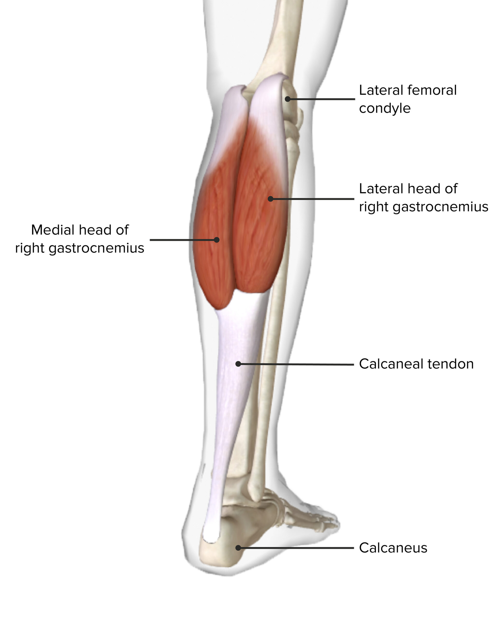
00:00 in the later lecture. Now let?s turn to the popliteal fossa. This is the important region directly behind the knee joint. And we can see on this diagram, we?ve got a quite intact popliteal fossa with the deep fascia still in place, and some cutaneous veins, the great saphenous and the short saphenous is here where the small saphenous sometimes labelled. And here when it?s been removed, we can see into the popliteal fossa and look at its boundaries and the contents. So the popliteal fossa is this fat-filled diamond-shaped space located posterior to the knee joint. It contains all of the neurovascular structures that pass from the thigh to the leg. Its boundaries are formed by the muscles that we mentioned previously. So superolaterally, here, we?re looking at the posterior surface of a right leg, right knee joint. So this is going to be a lateral aspect. This is going to be a medial aspect. Superolaterally, we have biceps femoris. Superomedially, we have semimembranosus just running down here. And this forms the top parts of our diamond. Inferolaterally, we have the lateral head of gastrocnemius, so down here. And inferomedially, we have the medial head of gastrocnemius which is running down here. We can see now we have the contents of the popliteal fossa where we?ve got the boundaries being formed by these muscles, so the contents within the popliteal fossa. If we look at the roof of the popliteal fossa, then it?s going to be popliteal fascia that the deep fascia lying over the popliteal fossa, and also the skin. And then superficial to that facia, we?ve got the great saphenous and the small or the short saphenous veins. The floor of the popliteal fossa is going to be the popliteal surface of the femur, and also a small muscle which we?ll see later on known as popliteus. And the contents, these structures here, are going to sit on that floor. So as I mentioned, the popliteal fossa contains all of the neurovascular
About the Lecture
The lecture Popliteal Fossa: Boundaries – Gluteal Region and Posterior Thigh by James Pickering, PhD is from the course Lower Limb Anatomy [Archive].
Included Quiz Questions
Which statement about the contents of the popliteal fossa is correct?
- It comprises fat and neurovasculature.
- It is mostly muscle.
- It is a sinus space.
- It is a blood-filled space.
- It is filled with tissue fluid only.
Which boundary of the popliteal fossa does the biceps femoris form?
- Superolateral
- Superomedial
- Inferomedial
- Inferolateral
- Posterior
Author of lecture Popliteal Fossa: Boundaries – Gluteal Region and Posterior Thigh

James Pickering, PhD
Customer reviews
5,0 of 5 stars
| 5 Stars |
|
5 |
| 4 Stars |
|
0 |
| 3 Stars |
|
0 |
| 2 Stars |
|
0 |
| 1 Star |
|
0 |
