Playlist
Show Playlist
Hide Playlist
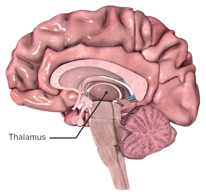
Thalamus

-
Slides 16 Diencephalon BrainAndNervousSystem.pdf
-
Download Lecture Overview
00:00 Next, I want you to understand a very important functional aspect of the thalamus. 00:08 The thalamus is a very important relay center for sensory information. All sensory information is relayed through thalamic connections up then to the cerebral cortex with the exception of olfaction. This slide is simpler in that it’s not overwhelmed with labels. 00:32 But it is demonstrating the thalamic radiations so these would be the connections that exist between the thalamic nuclei and the cerebral cortex. Thalamic fibers are shown in through here, these darker-colored structures. This is a more anterior view of those thalamic radiations. 00:52 This is a more posterior view of thalamic radiations. The thalamic radiations travel in the internal capsule. So they do accompany the motor axon fibers that are descending within the internal capsule. The motor fibers are shown here in the lighter color descending through the internal capsule. The thalamus may be considered the gate to consciousness. 01:25 Here, we see the oval thalamic nuclei. These are bilateral. Each one of these bilateral structures contains multiple nuclear components that make this a very complex structure. 01:44 Then here in the axial view, we can see various thalamic nuclei and their relationships to one another. I don’t mean to overwhelm you at the beginning here but the purpose of this slide is just to provide you a menu of the various thalamic nuclei. We are going to take a look at each one of these in greater detail, so I’m not going to read all these off to you at this moment. 02:17 But let’s now take a look at the anterior thalamic nucleus. The anterior thalamic nucleus is shown in through here and has been highlighted for you. When we think about thalamic nuclei, in this case the anterior one, we need to think about input, output. So what kind of information is coming in to this anterior thalamic nucleus? Well, the input here is from the mammillary body and the hippocampal formation. Now that the anterior thalamic nucleus has received this input, where is it going? What’s its output? Well, the output will be to the cingulate gyrus which is in the cerebral cortex. The function of your anterior thalamic nucleus is that it’s involved in the limbic pathway. The limbic pathway is discussed in detail in another lecture. 03:18 Next, we have the ventral posteromedial nucleus or simply VPM and it is highlighted here. 03:28 It too would have an input consideration and an output consideration. Input to the VPM is sensory from the face and taste, gustation. The output from this nucleus is going to be to the somatosensory cortex. Consequently, this nucleus is involved in relaying somatosensory cranial nerve inputs, cranial nerve inputs and taste or gustation to the cortex. We also have for your consideration, the ventral posterolateral nucleus of the thalamus, VPL and it is highlighted here. Its input is going to be sensory from the body and the limbs. This is going to process a lot of information coming into the thalamus. As it processes it, it’s going to output it, send it to the somatosensory cortex so we can perceive various sensations: touch, pressure, vibration for example. Again, it’s going to relay somatosensory spinal inputs up to the somatosensory cortex so that we can perceive those various senses. 04:58 Next on our list is the ventral anterior/lateral thalamic nuclei, the VA/L. These are highlighted in the image, so they can be seen here. Their input is going to be from the basal ganglia as well as from the cerebellum. Output from this collection of two nuclei is going to be to the motor, premotor, and supplemental motor cortices. These are located in the frontal lobe. 05:35 Consequently, this is the relay then between the basal ganglia and cerebellar inputs to the motor cortical areas. Another thalamic nucleus is the mediodorsal nucleus. 05:51 This is not shown in the plane of section that we have in the image. The inputs here are from the amygdala and olfactory inputs as well as limbic basal ganglia, so multiple inputs here. But the output is all to the frontal cortex. As a result, one of the functions here is for the limbic pathway then to allow a major relay to the frontal cortex allowing for that interconnectedness. This area can be damaged, can be lesioned and result in Wernicke–Korsakoff syndrome. Next is the pulvinar nucleus. The pulvinar nucleus is shown here in a light blue color. It’s receiving input from the visual pathway, auditory pathway and then there are other sensory pathways that will contribute input into this nucleus. Output is going to be to the parietal-temporal-occipital association area. This is going to allow us to orient ourselves toward visual, auditory, and other sensory stimuli. Here’s another thalamic nucleus that I want you to think about and remember. This is the medial geniculate body, MGB. 07:32 It’s labeled, a little slender portion of it right along in through here. The input into the medial geniculate body is going to be from the inferior colliculus. Its output is going to be to the auditory cortex of the temporal lobe. Thus, it functions as a relay for auditory input to the cortex. If we have a medial geniculate body, it stands to reason that we have a lateral geniculate body, LGB and that we can see it right in this particular area, not much there in the illustration or image. But this is the area that’s going to receive information from the retina, so that’s the input. Output then will be to your primary visual cortex in the occipital lobe. 08:32 So this is a very important relay nucleus then of visual input to the cortex for the perception of vision. This thalamic nuclear collection is the intralaminar/midline thalamic nuclei. 08:50 They are shown in this general area on the medial side, this collection of thalamic nuclei. 08:59 These are involved in arousal. As a result of that, they will activate the reticular, will be part of the activation of the reticular activating system. Here, we could focus on the lower portion of the slide. We’ll just come right in here to this center box. Right below it, you see various sensory inputs: vision, audition, olfaction for example. Let’s just say you have a visual stimulus. Now, let’s take a look at what happens next in response to the visual stimulus. 09:42 This will have excitatory output to the reticular activating system. We can see that excitatory input from the visual stimulus into the reticular activating system that is found in the brainstem. 09:58 From here, there is input that’s excitatory to thalamic nuclei. And so, we see the excitatory input to nonspecific thalamic nuclei. Then from here, you relay to the cortex to activate, to stimulate it. That’s the connectedness, the circuitry that exists between thalamic nuclei and the reticular activating system.
About the Lecture
The lecture Thalamus by Craig Canby, PhD is from the course Diencephalon. It contains the following chapters:
- Thalamus
- The Reticular Activating System
Included Quiz Questions
Which of the following thalamic nuclei gives its output to the primary visual cortex?
- Lateral geniculate body (LGB)
- Pulvinar
- Medial geniculate body (MGB)
- Ventral posterolateral (VPL)
- Mediodorsal
Which of the following thalamic nuclei, if destroyed, cause Wernicke-Korsakoff syndrome?
- Mediodorsal nucleus
- Lateral nucleus
- Midline nucleus
- Ventral posteromedial (VPM)
- Ventral posterolateral (VPL)
Which of the following thalamic nuclei relay sensory information coming from the body and lower limbs?
- Ventral posterolateral nucleus
- Anterior nucleus
- Midline nucleus
- Ventral posteromedial nucleus
- Mediodorsal nucleus
Which of the following thalamic nuclei helps us to orient toward visual, auditory, and sensory stimuli?
- Pulvinar
- Anterior
- Ventral posteromedial (VPM)
- Ventral posterolateral (VPL)
- Mediodorsal
Author of lecture Thalamus

Craig Canby, PhD
Customer reviews
5,0 of 5 stars
| 5 Stars |
|
5 |
| 4 Stars |
|
0 |
| 3 Stars |
|
0 |
| 2 Stars |
|
0 |
| 1 Star |
|
0 |
