Playlist
Show Playlist
Hide Playlist
Electron Microscopy


-
Slides GlomerularDiseaseOverview RenalPathology.pdf
-
Download Lecture Overview
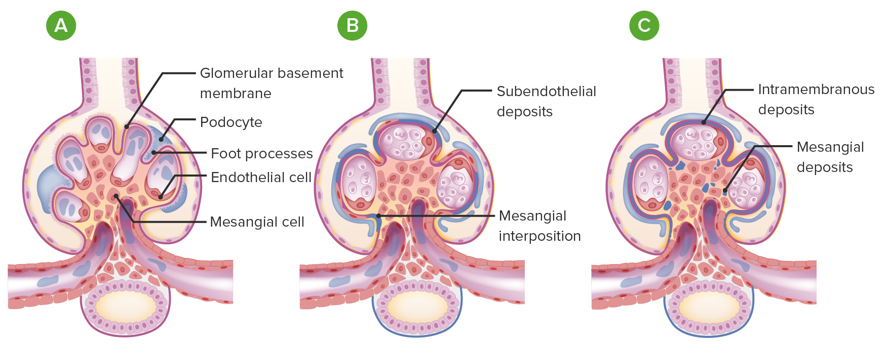
00:00 Let us now continue. 00:01 Now, electron microscopy is then going to help you identify few things. Light microscopy may have shown you or increased your suspicion of glomerular disease. You do electron microscopy. 00:15 What color would this be? Black and white. Fusion of your podocytes. Close your eyes. 00:22 Think about your podocytes. Where are you? You are on the side of the urine. You are on the other side of the glomerular basement membrane and that podocyte has foot processes in which they then become fused. Prototype, minimal changed disease. On electron microscopy, you actually find the fusion of the food processes. Such a finding would never take place at microscopy. 00:46 What about in immunofluorescence? What does immunofluorescence fluoresced or recognized? Immunoglobulins. 00:56 The fusion of foot processes has nothing to do with immunoglobulins, does it? No. So, therefore, why would you ever want to use immunofluorescence when you have fusion of podocytes? You don't. Use common sense. Know as to what information that you are getting. 01:12 Why it has been given? That it would then lead you into your diagnosis. Damage to what? Your visceral epithelial cell a.k.a. podocyte. What else? Well with electron microscopy, apart from the fusion of your podocytes, it detects IC, stands for immune complex. So, immune complex deposition, just takes us back to our previous discussion with immunofluorescence. 01:39 So, for example, let us say that electron microscopy did show you deposits, where? Maybe, on the side of the epithelial. Maybe, on the side of the endothelial. The electron microscopy shows you such dark deposits. Then on immunofluorescence what kind of pattern is this known as? Granular. 02:02 You go as far as that from now, for your boards and wards , you are in fantastic shape. 02:08 Now as you go in further specialization, let us say in pathology and understand there's certain techniques that you can use along with immunofluorescence that will then tell you if you are on the side of your endo or epithelial. The sites are designated as follows. Now pay attention here. Subendothelial. You are on the side of the blood. If you are underneath subendothelial, you will be between as the name says here or as the statements say here between the glomerular basement membrane and the endothelial cell. Is that clear? If it is isn't, I would recommen that you go back and take a look at the picture in which we walked through normal electron microscopy, identify glomerular basement membrane and the endothelial cell and what would you find in electron microscopy? A dark deposit. Now, what if you it did with the immunofluorescence? Is that even relevant? Of course, it is because it is immune complex. And here how would you then describe the pattern? Granular. Another one for electron mircroscopy. However, this will be subepithelial, so what are you under? Your podocyte and your glomerular basement membrane. This is once again an immune complex. On immunofluorescence, what kind of pattern is this called again? Very good, granular. Or you can have issues within the basement membrane. 03:39 For example, you have heard of membranous glomerulonephritis, haven’t you? MGN, membranous glomerulonephritis. What does that mean to you? Intramembranous issues, electron microscopy or maybe mesangial. Mesangial would be much better seen in terms of its changes if you did a light microscopy with H&E stain. The basis of what you need to know in terms of moving forward, laboratory investigations and biopsy is here in these discussions. 04:10 It is only once you have mastered the foundation of your investigation, you will then move into the particular pathologies. To summarize, three major biopsies stainings, we have our light microscopy H&E, we have immunoflurorescence, and number 3 electron microscopy. Let's move on.
About the Lecture
The lecture Electron Microscopy by Carlo Raj, MD is from the course Glomerulonephritis.
Included Quiz Questions
Which of the following is not detected by electron microscopy?
- Damage to the visceral endothelial cells.
- Fusion of podocytes.
- Damage to the visceral epithelial cells.
- The site of immunocomplex deposition.
- Fusion of visceral epithelial cells.
Which of the following is true regarding the site of depositions that pass through the glomerular basement membrane but are caught beneath the podocytes?
- Subepithelial
- Subendothelial
- Mesangial
- Epithelial
- Intramembranous
Which of the following deposition types can be much better visualized with light microscopy?
- Mesangial
- Subendothelial
- Endothelial
- Intramembranous
- Subepithelial
Author of lecture Electron Microscopy

Carlo Raj, MD
Customer reviews
5,0 of 5 stars
| 5 Stars |
|
5 |
| 4 Stars |
|
0 |
| 3 Stars |
|
0 |
| 2 Stars |
|
0 |
| 1 Star |
|
0 |
