Playlist
Show Playlist
Hide Playlist
Different Types of Bone

-
Slides 08 Types of Tissues Meyer.pdf
-
Download Lecture Overview
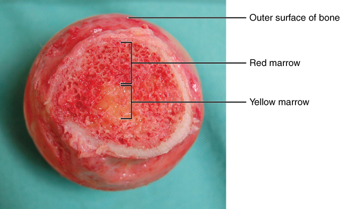
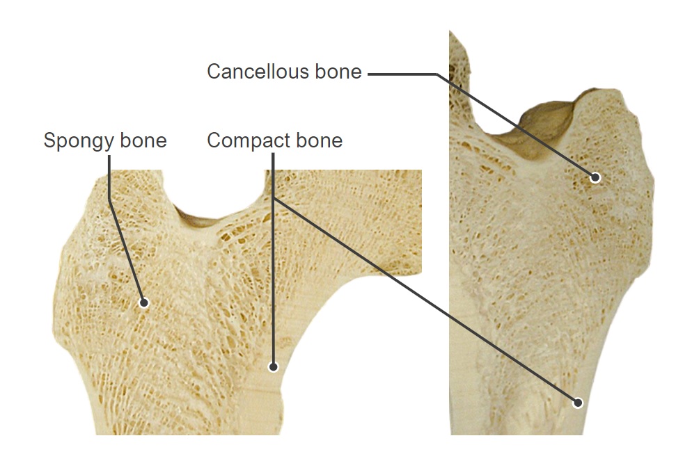
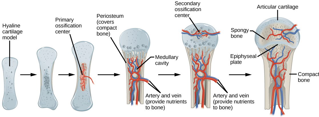
00:00 Again, like cartilage, the most important functional part of bone is the matrix. 00:02 Now, there are different types of bone. If we look at this image, this is a section through the very top part of the femur or the bone in our thigh that articulates with our hip joint and even at this gross level you can see that there is some area of the bone that is compact on the very outside of the bone. It is compact, very very hard. But towards the articular surface, the bone is very spongy. We call it spongy bone or we can also call it cancellous bone. And those little bony components you see within the cancellous bone or the spongy bone are often referred to as bony spicules. Bones are also classified as being long bones, short bones such as in the wrist, the hand joints, flat bones such as in the cranium or irregular bones that we also find in the wrist joint. These are gross anatomy classifications. Really what we want to concentrate today is the histological classification, and that is spongy bone or cancellous bone and compact bone. If you look very very carefully at this section of the spongy bone, you can see that the trabecula or the spicules of bone are all entitled in certain directions. The job of these spicules of bone is to impart the force of gravity and forces generated by movement down the shaft of the bone, down the compact part of the bone. And these can be rearranged. Bone is a very very active tissue. If you undergo weightlifting program or go into weightlessness in space, where the force is changed on the spicules of bone, then the bone changes the directions and the components of the spicules and the amount of the spicules to either withstand the increased load on the bone or if the load disappears, these spicules also disappear. But the key point is bone is a very very active tissue. Here is a structure of a long bone. 02:45 Again it is the bone in our thigh. It articulates with our pelvis at the hip joint. And really there are three or four major gross anatomy components of the bone. We call the shaft of the bone, the diaphysis. Towards the articular end of the bone, we call that part the epiphysis and the region just between the two in other words, the end of the shaft of the bone and the beginning of the epiphysis, we call the metaphysis. So these are gross anatomical definitions. But I want you to ask you to do something during this lecture. I want you to look at this bone and remember something forming. I want you to imagine that if you looked at this bone in real life, some of you may have looked at bones in the gross anatomy lab or other occasions. If you look very closely at this bone, you would see that there are some very very small holes in the bone. These very small holes represent where blood vessels enter the bone. I mentioned earlier that bone is very vascular. So I want you to imagine blood vessels coming into the bone at about the level of the diaphysis. 04:18 And I want you to imagine that these blood vessels travel up and down the compact bone that I pointed out earlier, and also travel into the internal part of the bone, which is hollow. 04:32 We call it the medullary cavity. And I want you to also imagine blood vessels passing up the compact bone and supplying the spongy bone, supplying that enormous surface area of bone with blood and therefore nutrients. So if you can imagine that, I am going to ask you to recall that imagination later on when we talk about how bone receives its nutrition. 05:04 Well, here is an H&E section taken of bone. It happens to be decalcified. All the matrix is taken out and we can see some of the organic components of the bone. And what I want you to focus on in this slide, are the images of the periosteum, the capsule around bone. Cartilage has a capsule or at least hyalin cartilage and elastic cartilage has a capsule, but that is called the perichondrium. 05:42 Well the periosteum has an outer fibrous layer, which is really just dense connective tissue. 05:52 But also lying very very close to the bone itself, is a very thin osteogenic layer. And that is a very important layer because that layer persists during our life. And if we break a bone, then that layer can be activated to help repair that break. On the outside part of both of these slides, both of these images, you can see skeletal muscle, and that skeletal muscle is going to insert into the bone via a tendon. Sometimes when you look into the bone matrix, you can see the osteocytes sitting in their lacuna. But also you can see these lacuna that seem to have dark pink contents. They are called Sharpey's fibres. 06:47 They are not cells. They are not the osteocytes, but they are in fact collagen bundles, collagen fibres that insert into the bone from the tendon. And of course that is very important because when the muscle contracts and the force of contraction is imparted through the tendon onto the bone by having this very strong cement or association with the bone, the tendon can then easily pass on that force of contraction onto the bone through the Sharpey's fibres and affect movement. Here is a ligament. In the middle of the section, we have the periodontal ligament. This periodontal ligament attaches the tooth to the bony socket, the alveolar bone and you can see in part where the collagen from the periodontal ligament inserts into the compact bone. Again these are called Sharpey's fibres. 07:56 Well let us go back to the section of the femur, the thigh bone. Try and point out yourself an area where this compact bone and try and point out to yourself where there is spongy or cancellous bone. But we really need to understand that the bone has always cavities inside them and surfaces and we really need to understand what covers these surfaces. At the very top, the articulating surface of bone has articular cartilage over it or hyalin cartilage, and this forms the articular joint. I have already spoken about periosteum, which forms the capsule around the bone. It ends of course at the articular cartilage because articulate cartilage does not have any periosteum around it nor is it covered by perichondrium. It is a friction free surface, lubricated by synovial fluid. Now within the bone, covering all those little spicules of bone that I described earlier, is the very very fine fine specialized connective tissue. 09:22 It is called endosteum. And I want you to remember that these endosteal cells are very important because again they can be stimulated to lay down bone, to repair bone, and they also have a very important function to do with nutrition of bone that I will talk to you about later on in this lecture. When you look at the diaphysis part of the long bone, it is hollow. 10:01 I have explained earlier that it has a medullary cavity within it. And during very early life, this medullary cavity and also within all the spaces between the spicules or trabecular bone in the spongy area, this area is dominated by red bone marrow, the haemopoietic organ that produces all our blood cells. And of course that will be the subject of a later lecture. 10:32 Once all our bone cells are formed, our bone marrow begins to start to disappear. And it only remain in certain parts of bones particularly in ribs. And when it disappears, as it does in the case of these long bones, it is replaced by fatty tissue and we call that yellow bone marrow. Because it has a yellow appearance, that fatty appearance.
About the Lecture
The lecture Different Types of Bone by Geoffrey Meyer, PhD is from the course Bone Tissue.
Included Quiz Questions
Which type of the following bone types is different from the other 3 types?
- Compact bone
- Spongy bone
- Trabecular bone
- Cancellous bone
Which statement is true about the endosteum?
- It supplies nutrients to the bone.
- It is a cancerous outgrowth.
- It is an outer layer of the bone.
- It has no useful functions.
- It promotes resorption of the bone.
Which of the following is another term for spongy bone?
- Cancellous bone
- Compact bone
- Cortical bone
- Lamellar bone
- Woven bone
Carpal bones are which of the following types of bones?
- Short bones
- Flat bones
- Sesamoid bones
- Regular bones
- Long bones
What section of a long bone contains the articular surface of the bone?
- Epiphysis
- Diaphysis
- Metaphysis
- Medullary cavity
- Periosteum
Which of the following is the osteogenic layer of the bone?
- The inner cellular layer of the periosteum
- The outer fibrous layer of the periosteum
- Articular cartilage
- Osteon
- Interstitial lamellae
The outer layer of the periosteum is composed of which of the following?
- Dense connective tissue
- Areolar connective tissue
- Loose connective tissue
- Epithelial tissue
- Lamellar bone
Which of the following is true about endosteal cells?
- They can lay down bone.
- They induce apoptosis of osteocytes.
- They have no function in adult humans.
- They are stem cells.
- They induce necrosis of bone.
Author of lecture Different Types of Bone

Geoffrey Meyer, PhD
Customer reviews
5,0 of 5 stars
| 5 Stars |
|
1 |
| 4 Stars |
|
0 |
| 3 Stars |
|
0 |
| 2 Stars |
|
0 |
| 1 Star |
|
0 |
Well, I find it more useful. I got what I want from this lecture.
