Playlist
Show Playlist
Hide Playlist
Pneumonia: Overview


-
Slides 01 URTIBronchitisPneumonia RespiratoryAdvanced.pdf
-
Download Lecture Overview
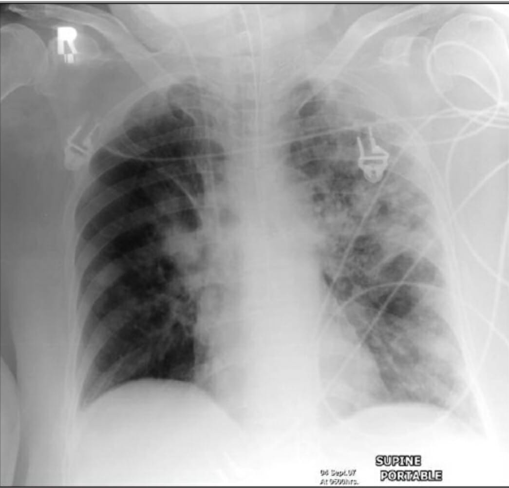
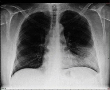
00:00 So moving on to discuss pneumonia. The hallmark, the defining situation with pneumonia is that you have infection of the alveoli and that causes consolidation. So that is where the air that’s in the alveoli has been replaced by an inflammatory exudate, bacteria, white blood cells, and red cells replacing the air, and you can see that on the chest X ray. So for example this chest X ray shows somebody with a right upper lobe pneumonia, and there is a white patch over the right hand of the lung, and that reflects the dense consolidation is that's occurring in that area, and there is no air left in those alveoli. And this distribution of pneumonia is a lobar pneumonia, it's affecting one lobe only and the rest of the lungs are clear. That’s the commonest presentation on an acute pneumonia. You can get pneumonia which is patchy throughout both lungs, so little patches throughout both lungs and that's called bronchopneumonia and this is an example of that. This is a much more serious disease in general because that’s a very extensive infection and is often a fatal event in patients with underlying severe disease such as a cancer or another disability such as dementia or chronic neurological deficit of some description. A third pattern of pneumonia that we can see is interstitial pneumonia. Now you probably can't tell very easily on this chest X ray that it's abnormal but there is a widespread reticular nodular infiltrate in both lungs, but it’s very subtle and that represents again a diffused pneumonia and is commonly associated with the atypical organisms that I mentioned earlier, Mycoplasma and Chlamydia. 01:44 The overall importance here is that, if you have pneumonia there is some form of alveolar consolidation, usually lobar but potentially interstitial or bronchopneumonia as well. 01:53 Now we can detect that clinically, so if you have somebody with pneumonia, the area of infected lung, the area of consolidation will be dull to percussion, and when you listen to it they'll be crackles in that area, and potentially bronchial breathing. And the chest X ray as we’ve seen is obviously abnormal. It shows airspace shadowing with white patches in the infected area, and occasionally what we call air bronchograms. That’s where the bronchus that’s moving through the consolidated lung has been delineated by the alveolar consolidation I've stated here. So it shows up as a black tube going through a consolidated area.
About the Lecture
The lecture Pneumonia: Overview by Jeremy Brown, PhD, MRCP(UK), MBBS is from the course Infections of the Respiratory Tract.
Included Quiz Questions
Which of the following is NOT a component of the inflammatory exudate that replaces air in a pneumonic consolidation?
- Hyaline membranes
- White blood cells
- Red blood cells
- Bacteria
Which of the following is TRUE regarding pneumonia?
- Bronchopneumonia presents as patchy infiltrates on X-ray.
- Lobar pneumonia is more fatal in comparison to bronchopneumonia.
- Lobar pneumonia involves all lobes of the lungs.
- Lobar pneumonia is usually caused by atypical organisms.
- Interstitial pneumonia is the most common type of pneumonia.
Author of lecture Pneumonia: Overview

Jeremy Brown, PhD, MRCP(UK), MBBS
Customer reviews
5,0 of 5 stars
| 5 Stars |
|
5 |
| 4 Stars |
|
0 |
| 3 Stars |
|
0 |
| 2 Stars |
|
0 |
| 1 Star |
|
0 |
