Playlist
Show Playlist
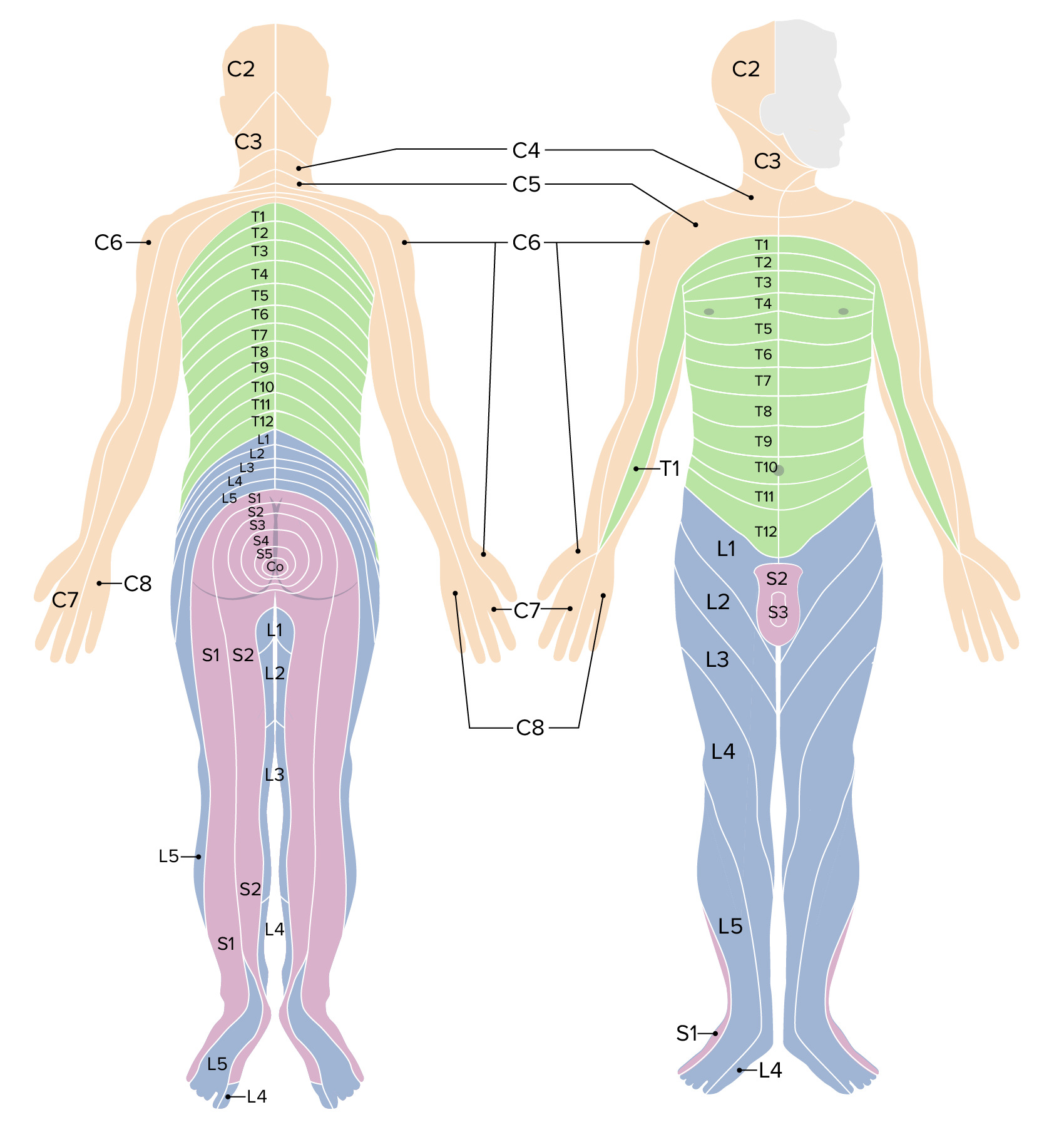
Hide Playlist
Nervous System: Structure and Function of the Peripheral Nervous System – Biological Bases of Behavior (PSY, BIO)

-
Slides Biological Basis of Behavior.pdf
-
Download Lecture Overview
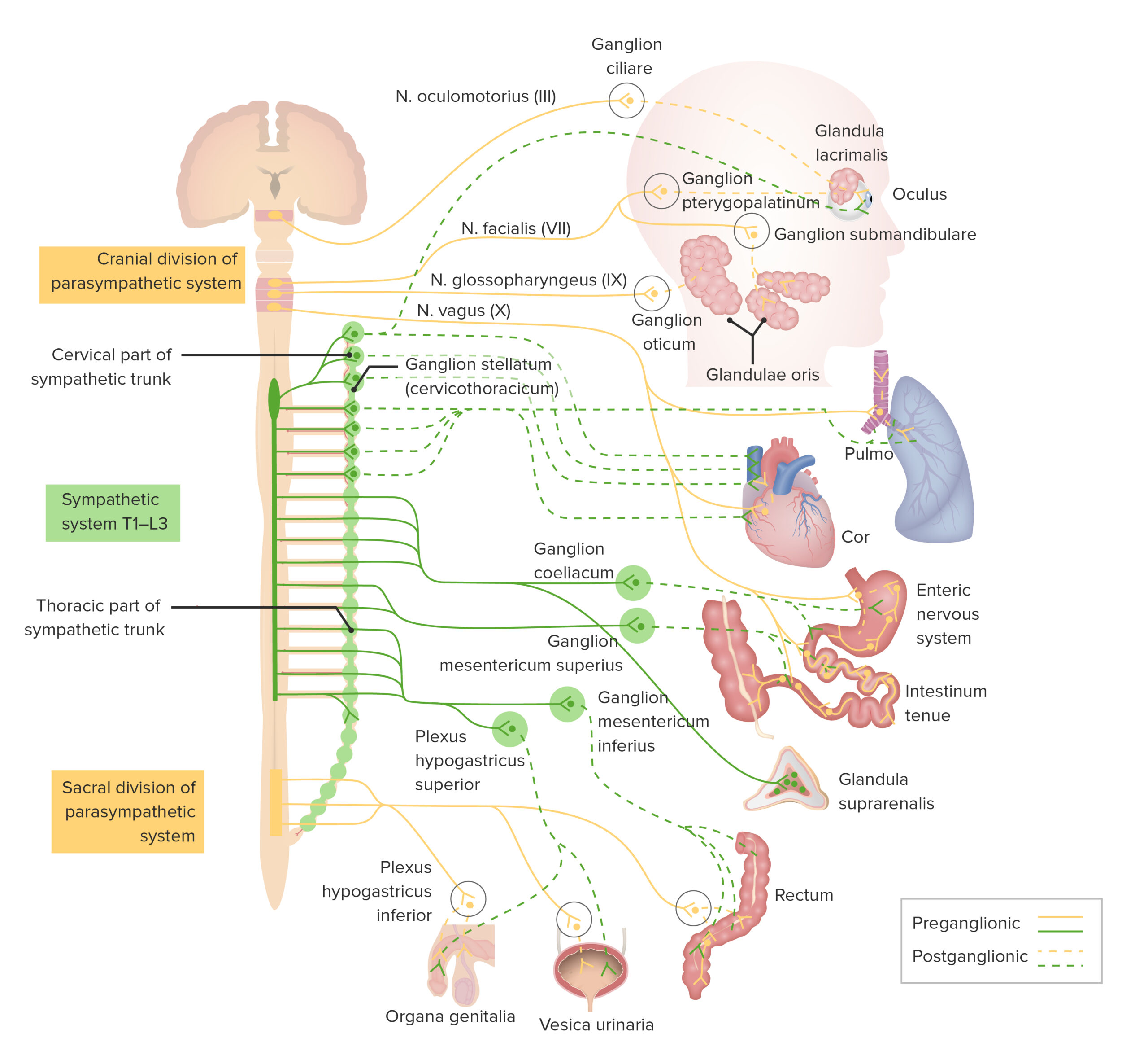
00:01 Okay. So now, we’re going to look at that -- we’re going to continue to look at some of that organization of the nervous system. 00:05 So, very quickly we have two bins, the CNS, the PNS, central nervous system, peripheral nervous system. 00:11 This is not new terms for you, I’m sure. 00:13 And I’m sure you’re familiar with what they represent. 00:16 Simply put the central nervous system is the brain and spinal cord and the peripheral nervous system is everything else. 00:22 So it’s referring to all the nerves and the sensory structures outside of the brain and spinal cord. 00:27 So, we can break down the PNS a little bit further. 00:29 We have the somatic system and the autonomic system with the autonomic -- the autonomic containing the involuntary control of glands and smooth muscle. 00:37 And the name autonomic refers to the fact that it’s essentially automatic. 00:41 So you’re not sitting there right now thinking, “I need to think about stomach digesting or I need to think about breathing.” It just happens, right? So that means it’s done automatically and this is all involuntary control. 00:54 It includes the sympathetic and parasympathetic systems, which is the fight or flight or rest and digest, and we’ve talked about those quite a bit. 01:02 And you recall from the previous or upcoming lectures, fight or flight is in response to a stimulus. 01:09 Are you going to stay there and address the stimulus that’s in front of you fight or are you going to turn around and run away. 01:15 So the analogy that’s always used is caveman days being presented with a tiger or a, you know, something else. 01:21 So saber-toothed tiger is about to attack you, are you going to stay and fight, or are you going to turn around and run back to your cave? And that’s an automatic response that happens quite quickly. 01:30 And it happens to you I’m sure. 01:32 Same thing, maybe not a saber-toothed tiger but if confronted with a threat, do you fight, do you address it, or do you turn around and run away? And then there’s the parasympathetic which is the rest and digest. 01:43 Again, we’ve talked about that in quite a bit of detail. 01:47 So, let’s take a look at how the PNS is anatomically organized. 01:52 So, all the neurons entering and exiting the central nervous system are carried by sort of two major categories here, cranial nerves and spinal nerves. 02:01 So, there are 12 pairs of cranial nerves that convey sensory and motor information to and from the brainstem which is from down below. 02:09 And then we have 31 pairs of spinal nerves which convey sensory information and motor information to and from the spinal cord. 02:16 So we have two broadband. 02:17 We should be pretty familiar with that. 02:19 And here’s an example of the vagus nerve. 02:21 So the vagus nerve is an example of a cranial nerve and it decreases heart rate and increases GI function. 02:26 And you can see how sort of convoluted it is and it’s going through -- it’s part of the parasympathetic nervous system -- sorry, parasympathetic division of the autonomic nervous system and it travels up to the brain. 02:38 Okay? So, the point is, you have a lot of info coming from outside and it goes via these two means to those two different spots, so the brainstem or the spinal cord. 02:48 Now, we’re going to compare sort of somatic motor neurons versus sensory neurons. 02:54 Now, the motor neurons, they innervate skeletal muscle cells and they use acetylcholine as their primary neurotransmitters. 03:01 And the cell bodies are in the brain stem or the ventral front portion of the spinal cord. 03:05 Now, on the other hand, the sensory neurons have a long dendrite which extends from a sensory receptor in the skin or muscle, so anywhere outside to bring information in and it’s connected to the soma or cell body through the -- the DRG or the dorsal root ganglion. 03:21 Again, something that we’ve seen before. 03:24 There’s a pair of DRG for every segment on the spinal cord. 03:27 And these are protected by the vertebral column but their outside of the meninges. 03:32 And we’ll take a blowup of the spinal cord in just a moment. 03:36 But the point is, they’re outside of the central nervous system. 03:39 Okay? So, sensory neurons bringing information -- sensory information to the brain via the DRGs and it does so through the spine. 03:48 And we have our motor neurons which bring -- sorry innervate muscle. 03:54 Okay? So we have that -- those two modes of information. 03:59 Now, the autonomic peripheral nervous system is organized in a certain fashion, and we have information going in and out. 04:07 So, all efferents of the sympathetic and parasympathetic system consist of two types of neurons. 04:13 Preganglionic neuron which has a cell body in the brainstem or spinal cord and postganglionic, this sends an axon to the effector muscle. 04:20 So in English, we’re basically saying we have -- we sort of have a connection with the preganglionic being stuff coming from the brain going out. 04:29 And then your postganglionic is a stuff that’s going to the actual intended target, so the muscle that you’re trying to move or the organ and gland that you’re trying to activate. 04:38 All autonomic preganglionic neurons release acetylcholine as their primary neurotransmitter. 04:43 So acetylcholine is extremely important when we’re talking about this type of peripheral nervous system activation. 04:50 All parasympathetic postganglionic neurons also release acetylcholine. 04:55 Okay? So there are lots of terms that we’re throwing, preganglionic, parasympathetic, autonomic, and it might be kind of hard to grasp. 05:02 So one way to get through this is to have a little diagram, that flowchart that we showed, and it allows you to understand parasympathetic versus autonomic and what the roles are and preganglionic versus postganglionic, same thing, understanding which is what and then motor versus sensory information. 05:20 So, nearly all the sympathetic postganglionic neurons release norepinephrine as their neurotransmitter. 05:26 So, the primary players here are acetylcholine and norepinephrine between these two systems. 05:33 So let’s look at this through diagrams because pictures are always easier to grasp. 05:37 So, on the top, we have our somatic efferent innervation. 05:40 And then below, we have our autonomic efferent innervation. 05:43 So, somatic means that this is something that we are conscious about and we’re actually -- we’re sending a signal out to say your muscle to flex. 05:53 So you can see that a signal comes from the spinal cord through the ventral horn and it’s going to travel on the length of an axon, and then it’s going to release acetylcholine that’s going to activate that muscle. 06:04 And down below, we have the autonomic efferent innervation. 06:08 So stuff that like your heart beating. 06:10 Again, this isn’t something you’re thinking about, it just happens. 06:13 And you see the signal being passed, acetylcholine being released. 06:17 Now, we have the ganglion cells and then that’s where the synapse happens. 06:21 And then the norepinephrine or acetylcholine goes on and sent to the heart. 06:26 Okay, so two different processes. 06:28 There are some commonalities of course but you can notice the difference in transmitter and you can notice the autonomic ganglion cells. 06:37 Now, let’s talk a little bit more about the autonomic PNS anatomy. 06:44 All sympathetic preganglionic efferent, so things going out have their cell bodies in the thoracic or lumbar regions of the spinal cord. 06:53 So the spinal cord is a really unique, basically, highway that’s sending information up to the brain and sending signals out. 07:00 And it has a very important protective function as well. 07:04 So remember, the CNS is spinal cord and brain and PNS is everything else. 07:09 So this is why the sympathetic system is sometimes referred to as the thoraco-lumbar system because we’re involving these two components. 07:19 Now, the parasympathetic preganglionic efferent neurons have their cell bodies in the brainstem of the lower portion of the spinal cord or the sacral portion, which is why sometimes get that name craniosacral system. 07:31 In the preganglionic axons for the parasympathetic versus sympathetic system differ in length and we’re going to compare those two in a summary table in an upcoming slide. 07:42 Okay. So, let’s look at this overly complicated figure. 07:45 So I don’t want you to get frightened when you see this. 07:47 There’s a lot of information here. 07:49 And what this is, is an overview of sort of all the components, structure and function of the peripheral nervous system. 07:56 So what I really want you to get out of this is we’ve separated both the sympathetic and the parasympathetic system. 08:03 So, we’re looking at stuff that is sort of, you know, like the heart rate breathing versus organs and muscle control. 08:09 And you can see from the diagram different types of inputs, and that’s kind of really all that I want you to get to. 08:14 There are the two different systems and we have all the different organs, effectors, and you see the different input and how it’s going to go -- how it interjects into the spinal cord. 08:25 So you can see how the sympathetic stuff is going. 08:28 All the inputs are going up through the spinal cord as opposed to the stuff that is going to the brainstem. 08:34 So, you don’t need to necessarily memorize every single aspect of this diagram, but it’s just kind of strengthening some of the points that we’ve already mentioned. 08:42 Now, here’s a summary table that should hopefully help, sympathetic versus parasympathetic. 08:47 So the general function is the fight or flight for sympathetic that we mentioned, mobilized energy versus rest and digest is where relaxing, taking it easy. 08:56 Location of the preganglionic soma, it’s the thoracic and lumbar spinal cord for the sympathetic versus the brainstem for the parasympathetic. 09:04 In terms of the length of the axon: short versus long. 09:09 And then you can imagine why because for the sympathetic it’s going just to the spinal cord, whereas for the parasympathetic it needs to go up to the brainstem. 09:17 And then we have the ganglia, we say close to cord, far from target versus far from cord, close to target. 09:24 Okay? And then we have the postganglionic axon which is long and -- versus short. 09:29 And the transmitters involved here for the sympathetic are norepinephrine versus acetylcholine. 09:34 This is a nice kind of summary table. 09:36 This should definitely help you to put all those pieces together.
About the Lecture
The lecture Nervous System: Structure and Function of the Peripheral Nervous System – Biological Bases of Behavior (PSY, BIO) by Tarry Ahuja, PhD is from the course Individual Influences on Behavior. It contains the following chapters:
- Organization of the Nervous System
- Sympathetic vs. Parasympathetic Nervous System
Included Quiz Questions
Which neuron does not use ACh as a neurotransmitter?
- Sympathetic postganglionic
- Parasympathetic preganglionic
- Somatic efferent
- Sympathetic preganglionic
- Parasympathetic postganglionic
Which neuron is responsible for sending signals from thermoreceptors?
- Somatic sensory
- Somatic efferent
- Sympathetic preganglionic
- Somatic special sensory
- Parasympathetic postganglionic
Where are the cell bodies of sympathetic preganglionic neurons located?
- Lumbar spinal cord
- Coccygeal spinal cord
- Cervical spinal cord
- Brainstem
- Cerebral cortex
Where are the cell bodies of parasympathetic preganglionic neurons located?
- Sacral spinal cord
- Thoracic spinal cord
- Lumbar spinal cord
- Thalamus
- Cervical spinal cord
Which of the following is true regarding the axonal length of pre- and postganglionic neurons of the sympathetic nervous system?
- Preganglionic neurons are short and postganglionic neurons are long.
- Both preganglionic and postganglionic neurons are short.
- Both preganglionic and postganglionic neurons are long.
- Preganglionic neurons are long and postganglionic neurons are short.
- They are the same length as neurons of the parasympathetic nervous system.
Author of lecture Nervous System: Structure and Function of the Peripheral Nervous System – Biological Bases of Behavior (PSY, BIO)

Tarry Ahuja, PhD
Customer reviews
5,0 of 5 stars
| 5 Stars |
|
1 |
| 4 Stars |
|
0 |
| 3 Stars |
|
0 |
| 2 Stars |
|
0 |
| 1 Star |
|
0 |
All information is carefully thought out and clear! As a student who is starting to learn about behavioral science, these lectures are perfect for me!
