Playlist
Show Playlist
Hide Playlist
Clubfoot (Talipes Equinovarus) and Metatarsus Adductus

-
Slides DevelopmentalDysplasiaoftheHip Pediatrics.pdf
-
Download Lecture Overview
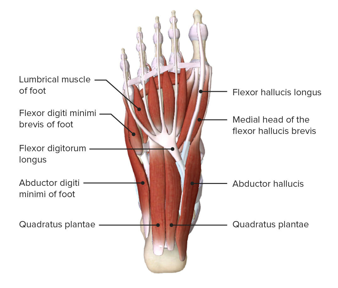
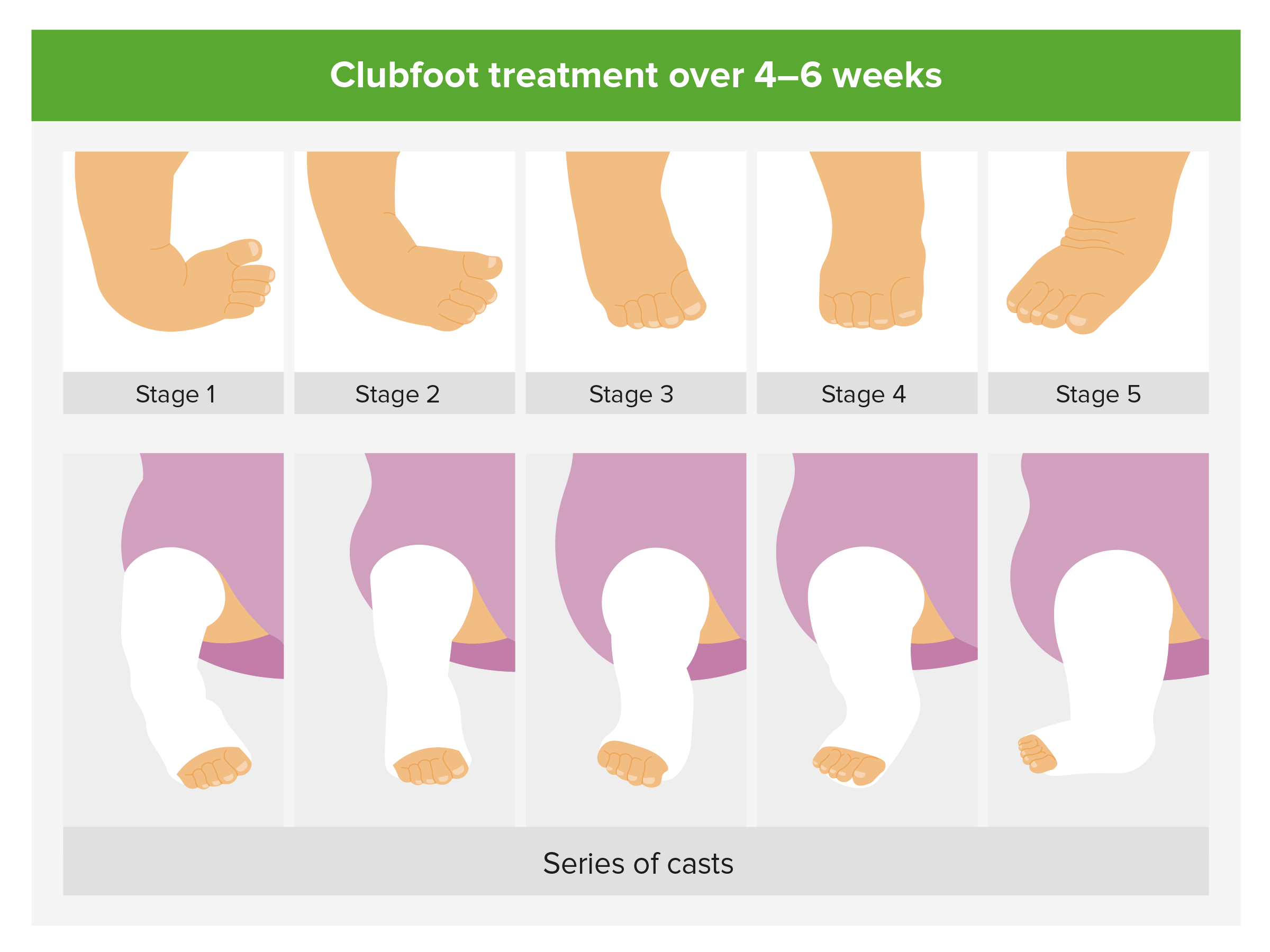
00:01 Let’s switch gears from DDH and start talking a little bit about clubfeet. The real name for clubfeet is talipes equinovarus. But I’m going to call it clubfoot for short. This is a congenital complex fixed foot deformity. Basically, the mid and forefoot are adducted. There is equinovarus of the hind foot. 00:26 This results in a high foot arch. Let me show you what I’m talking about. It’s generally idiopathic. 00:33 It may be associated with other problems such as developmental dysplasia of the hip or arthrogryposis. 00:41 There is a strong familial tendency even in distant relatives. We don’t know why but if great grandma has a clubfoot, her child, her great grandchild may as well. So, here’s a picture of a child with a clubfoot. 00:58 As you can see, this child will have thin calf muscles from underuse. The forefoot is adducted. 01:06 There is a shortened foot. The entire foot is inverted and supinated with the heel rotated inwards which is varus and the ankle in plantar flexion which is quinus. Here is a photo to show you what I’m talking about. It’s a shortened foot. It’s bent in and up, supinated as it were. 01:30 Basically, with a child who has a clubfoot, it’s noticed usually right at birth. Those children should be referred to the pediatric orthopedist. Left untreated, this child will start to learn to walk on the side or top of the foot and it will result in a lifelong limp. Initial treatment is usually with a variety of splinting and casting, a serial and gradual remanipulation of the foot into a correct position. 01:59 In some cases however, surgery is absolutely required. Usually around 6-12 months old after the patient starts a splinting and casting regimen, the child may eventually go to surgery. 02:13 If the child has a poor response to both splinting and casting and surgery, they may have further surgical correction at 5-6 years of age to continue to try and improve the function of that foot. 02:28 Let’s switch gears one more time to metatarsus adductus. This is also called metatarsus varus. 02:37 This is a deformity of the foot where the foot is turned inward as you can see in this picture. 02:42 This is the most common foot deformity in infants. About half the time, it’s bilateral. There is often a family history. It’s also associated with torticollis or developmental dysplasia of the hip, in other words, a tight uterine environment. The child has perhaps less hydramnios, oligohydramnios and that cramped uterine environment has caused this feet to become this way. So, what you will note on exam when you’re looking from the bottom of the patient is a medial deviation of the forefoot but a normal hindfoot. The forefoot can be possibly stretched into a neutral position. 03:24 Ankle flexion and extension is normal. Therapy is through passive stretching or you can do no therapy at all. This often will self-correct on its own by one to two years of age. 03:39 We do refer to orthopedics if it’s a fixed deformity, if it’s painful, or if the child is having difficulty finding shoes that work for that child. Let’s look now at intoeing in children, reasons why children tend to have their feet pointing in. This is a common complaint among parents coming to their primary care provider. There are a couple of different reasons why this happens. 04:07 One possibility is tibial torsion. The femurs and knees are facing forward but the tibiae have torsed inward. 04:15 This often occurs in toddlers and may be associated with bowed legs which is also called tibia varus. 04:21 Usually in children with intoeing, no therapy is needed. Just let the families know this will likely resolve in time. Patients may also develop femoral anteversion as a cause of intoeing. 04:38 Here, the knees are inwardly rotated because the problem is actually at the femoral insertion into the pelvis. The femurs themselves are anteverted or turned in. We see this commonly in children who like to sit in this sort of W position. Children sitting in this W position have an easy time internally rotating their femurs. Sometimes this is associated with joint hypermobility as in patients with Ehlers–Danlos syndrome. The good news is just like in tibial anteversion, this problem self-corrects. Usually it self-corrects by around eight years of age. So, that's a brief summary of the most common problems with the development of the legs in children. Thanks for your attention.
About the Lecture
The lecture Clubfoot (Talipes Equinovarus) and Metatarsus Adductus by Brian Alverson, MD is from the course Pediatric Rheumatology and Orthopedics. It contains the following chapters:
- Clubfoot
- In-toeing
Included Quiz Questions
Which of the following is TRUE about metatarsus adductus?
- 50% of cases have bilateral foot involvement.
- It is also called “tabes dorsalis”.
- It usually requires surgery.
- Family history for the problem is rare.
- It is associated with polyhydramnios.
Which of the following statements about Talipes equinovarus is CORRECT?
- It involves the adduction of the forefoot.
- It results in a low foot arch.
- It is a variation of the clubfoot.
- It is not related to developmental dysplasia of the hip.
- It is seen only in girls.
Which of the following is NOT a feature of clubfoot?
- Outward rotation of the heel
- Plantar flexion (quinus)
- Foot inversion and supination
- Short foot
- Thin calf muscles
A 6-month-old girl is diagnosed with clubfoot. Which of the following is the next best step in management?
- Splinting/casting
- Reassurance
- Pharmacotherapy
- Surgery
- Pavlik harness
What is the deformity seen in metatarsus adductus?
- Medial deviation of the forefoot.
- Medial deviation of the hindfoot.
- High arch of the foot.
- Inability to stretch the forefoot to the neutral position.
- Eversion of the forefoot.
Which of the following statements is FALSE regarding femoral anteversion?
- This results in fixed flexion of the knees.
- It is often seen in children who sit in a "W" position.
- It may indicate joint hypermobility.
- It usually self-corrects by 8 years of age.
- It is one of the causes of in-toeing in children.
Author of lecture Clubfoot (Talipes Equinovarus) and Metatarsus Adductus

Brian Alverson, MD
Customer reviews
5,0 of 5 stars
| 5 Stars |
|
1 |
| 4 Stars |
|
0 |
| 3 Stars |
|
0 |
| 2 Stars |
|
0 |
| 1 Star |
|
0 |
Excellent lecture. This is one of the key general pediatrics topic. So it's important to have some knowledge about it. Thank you.
