Playlist
Show Playlist
Hide Playlist
Malignant Liver Lesions

-
Slides Malignant Liver Lesions.pdf
-
Download Lecture Overview
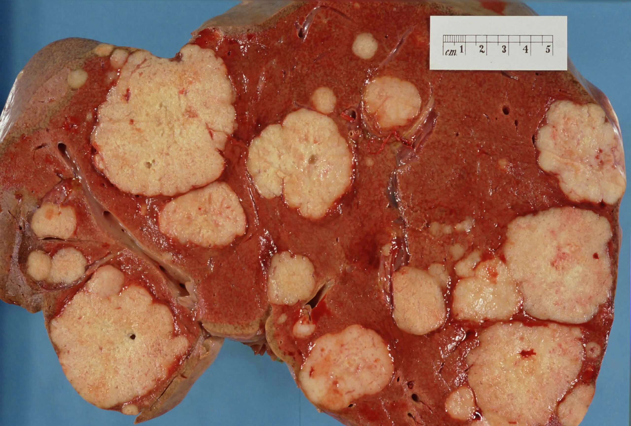
00:00 So in this lecture we?ll be discussing some common malignant lesions that can be found within the liver. 00:06 Let?s take a look at these three CT images. 00:10 Keep these three images in mind as we go through this lecture. 00:14 So the 2 most common liver malignancies are metastases and hepatocelular carcinoma. 00:22 Metastases are actually the most common malignancy found within the liver. 00:27 If you take a look at this pathological specimen you can see multiple masses throughout the liver and these represent metastases. 00:34 The most common organ of origin is the GI tract, particularly the colon. 00:39 Other common sites of origin include the breast, lung, and melanoma. 00:44 So how can you recognize a metastases on an imaging study? Usually, they come up as multiple low to intermediate density masses with slightly ill-defined margins. 00:56 They also demonstrate very variable enhancement after contrast administration. 01:00 Contrast-enhanced MRI and CT are about equally sensitive in the detection of metastases within the liver. 01:07 So let?s take a look at this CT scan. This is a non-contrast enhanced CT and it demonstrates multiple calcified metastases within the liver from a colon adenocarcinoma. 01:20 On the same patient, when you give contrast the contrast-enhanced CT actually shows multiple other masses that now demonstrate heterogeneous enhancement. 01:30 Let?s take a better look at this. So this is the non-contrast enhanced CT, you can see a couple of different masses with calcifications within it. 01:38 The high density here is calcification as we have not yet given contrast. 01:42 On this contrast-enhanced study you can see multiple, multiple, masses a lot more than you can see on the non-contrast examination and many of them demonstrate rim enhancement, some of them demonstrate central enhancement, so very variable enhancement actually. 01:56 Hepatocellular carcinoma is the second most common malignancy found within the liver. 02:00 About 80% of hepatocellular carcinoma cases are associated with either hepatic cirrhosis or hepatitis B or C. 02:08 Usually, hepatocellular carcinoma presents as a solitary mass, although occasionally it may present as multiple masses. 02:16 So this is an example of a CT demonstrating hepatocellular carcinoma. 02:21 This is a coronal arterial phase CT which demonstrates a heterogeneously enhancing mass that has prominent vascularity. 02:29 This entire portion of the liver looks like it?s abnormal and you can see multiple vessels within it. 02:35 On the portal venous phase you have washout of the enhancement, so now you see a very large mass with very little enhancement left within it. 02:46 And this is very typical of a hepatocellular carcinoma. 02:51 It demonstrates arterial phase enhancement and then washout in the portal venous phase, so early washout. 02:57 Most often a hepatocellular carcinoma presents as a hypodense or isodense lesion on a non-contrast CT so this can be a very vague finding. 03:08 Early enhancement in the arterial phase is seen as we just saw and we can have early washout on the portal venous and delayed phases. 03:16 Hepatocellular carcinomas can occur anywhere within the liver. 03:19 An MRI is actually a lot more sensitive as there are certain features that are really seen only on MRI and not really seen on CT. 03:25 So what are the MRI features of hepatocellular carcinoma? It usually presents as a hypointense mass on T1 weighted images. 03:35 It demonstrate mild hyperintensity on the T2 weighted images and may contain cystic or necrotic areas that are seen on T1 and T2 images and they are T1 hypointense and T2 hyperintense so it presents us a very heterogeneous mass even on MRI. 03:53 Post-contrast images demonstrates heterogeneous enhancement as they do on the CT that we just saw. 03:59 Usually, a biopsy has to be performed in order to really prove what the lesion is so a definite diagnosis really can?t be made just by imaging. 04:09 It really does need a biopsy and biopsies can be performed using either ultrasound or a CT guidance. So let?s take a look at this case. 04:19 There?s a liver lesion on the portal venous phase. 04:22 Can you describe what the findings are? Let?s take a look at the lesion and let?s take a look at everything else that?s on this exam that might help us better characterize with this lesion represents. 04:39 So there?s a low density lesion within the right lobe of the liver, we can see it right here relatively well circumscribed. 04:46 Associated findings include ascites so there is a lot of fluid surrounding the liver here that?s low density and you can see nodularity of the liver contour. 04:55 There?s a little bit of fluid around the spleen as well here. 05:00 So given the associated findings of hepatic cirrhosis, this lesion most likely represents a hepatocellular carcinoma and as you remember on the portal venous phase the lesion washes out and may not demonstrate enhancement. 05:15 So let?s go back to that initial case that we saw when we began the lecture. 05:21 So what CT phases are depicted here? What do you think the imaging findings are? So the first is the non-contrast enhanced CT so there?s no contrast administered on this one and you can see that there is a low density lesion near the hilum of the liver. 05:45 The second is an arterial phase which demonstrates an enhancing mass also at the hilum of the liver and on the portal venous phase, the mass demonstrates washout so very little enhancement is left here. 05:59 This actually represents a metastases. 06:03 It?s a low density, slightly irregular mass seen on a non-contrast CT image. 06:08 Again, the arterial phase demonstrates homogeneous enhancement and it actually has what we call a central nonenhancing nidus. 06:14 So the central portion of this mass is not enhancing the way the rest of the mass is enhancing. 06:20 In the portal venous phase you have washout of the contrast. 06:24 So although this could represent a hepatocellular carcinoma with the early enhancement and the early washout, homogeneous enhancement in the central nidus are not really typical for hepatocellular carcinoma so this most likely represents a metastases. 06:38 Again, this is one of those cases that we really can?t say a definite diagnosis of just basing on the imaging. The patient really would need a biopsy and this can be performed again either by ultrasound or CT guidance. 06:50 This patient actually has a known GI stromal tumor and based on that fact we can more definitely say that this likely represents a metastases. 07:00 So we?ve gone over some of the more common malignant liver lesions. 07:05 The 2 most common again are hepatocellular carcinoma and metastases.
About the Lecture
The lecture Malignant Liver Lesions by Hetal Verma, MD is from the course Abdominal Radiology. It contains the following chapters:
- Metastases and Hepatocellular Carcinoma
- Liver Lesions: Case Study
Included Quiz Questions
Which of the following Imaging findings are seen with hepatocellular carcinoma?
- Early enhancement and early washout
- Hyperdensity is seen on non-contrast CT
- Early enhancement and delayed washout
- Delayed enhancement and early washout
- Delayed enhancement and delayed washout
Which of the following is the MOST common malignancy found within the liver parenchyma?
- Metastasis
- Adenoma
- Hepatocellular carcinoma
- Choriocarcinoma
- Cholangiocarcinoma
How does metastasis in the liver from colon adenocarcinoma appear on a non-contrast enhanced CT?
- Multiple calcified lesions
- Hyperdense linear densities in the periphery
- Well-defined hypodense mass
- Laminated densities in the center of the liver
- A cystic lesion in the right lobe
Which of the following is FALSE regarding hepatocellular carcinoma?
- It does not have arterial phase enhancement on CT.
- It demonstrates heterogeneous enhancement on post-contrast images.
- About 80% of cases are associated with hepatic cirrhosis or hepatitis B or C.
- It is often solitary, although there may be multiple masses.
- Arterial phase CT demonstrates a heterogeneously enhanced mass with prominent vascularity.
Author of lecture Malignant Liver Lesions

Hetal Verma, MD
Customer reviews
5,0 of 5 stars
| 5 Stars |
|
5 |
| 4 Stars |
|
0 |
| 3 Stars |
|
0 |
| 2 Stars |
|
0 |
| 1 Star |
|
0 |
