Playlist
Show Playlist
Hide Playlist
Fascial Spaces and Surgical Access to Trachea – Neck

-
Slides 25 Neck1 HeadNeckAnatomy.pdf
-
Download Lecture Overview
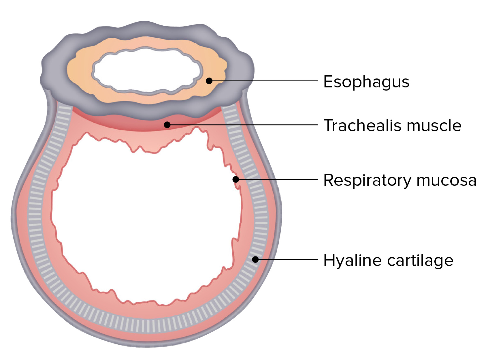
00:01 Now, we’re going to transition over to fascial spaces. There are three spaces that can form between the deep fascial layers. We’re going to be able to see two of these areas very, very well. 00:17 The third unfortunately is not well-illustrated in the image but we’ll make note of the fact that it does exist. First, we have the pretracheal space. The pretracheal space is shown anterior to the visceral compartment. That’s why it’s anterior to the trachea in this particular view. 00:44 A second space is shown right in through here. This is referred to as the retropharyngeal space. 00:56 Then it lies between the visceral compartment and its sheath and the vertebral compartment and its deep fascial layer. The third space is the prevertebral space. That space would be the deep fascial layer that exists over the anterior portion of the body of related vertebra and the anterior part of its transverse process. This is a bilaminar area so there’s a space between it. 01:31 Unfortunately, it is not well-illustrated in this particular image but it is one to keep in mind. 01:39 Why are these fascial spaces important? Well, one area of clinical correlation or relevance here is that infections and we’ll use the pretracheal space right in through here, if there’s an infection in this area, it may extend inferiorly and reach the anterior mediastinum. 02:05 A second clinical consideration is if invasion occurs, that is invasion of a cancer, the cancer can spread into any one of these three potential fascial spaces. A third and final consideration here is with respect to the retropharyngeal space, this space right in through here. If there’s an infection here, this can spread inferiorly and in fact to the posterior mediastinum and that could then form an abscess in this particular retropharyngeal space as a result. 02:53 Our next area to take a look at is surgical access to the trachea. This may be necessitated when one cannot intubate and you need to provide ventilation to an unconscious patient or even a conscious patient. Several approaches, one is a coniotomy. A coniotomy goes by two alternate names, a cricothyrotomy. This can also be referred to simply as a crike. 03:28 Another approach is to perform an upper tracheotomy which will be in this region here. 03:38 Then the third approach would be to do a lower tracheotomy shown in this region here. 03:45 We will now explore each one of these with the approach taken. Here, I’ll begin with the coniotomy or simply the crike. The relevant anatomy is the area between the inferior portion of the thyroid cartilage of the larynx that we see here. This laryngeal cartilage that we see inferior to the thyroid cartilage is the cricoid cartilage and would be right up here along its superior border. Running then between these two laryngeal cartilaginous structures is the cricothyroid membrane. One would get to this level surgically and then horizontal incision would be performed. Then the patient could be ventilated. This area here in a coniotomy is immediately inferior to the local cords or folds. The other approach is the tracheotomy. There’s an upper one and a lower one. Let’s take a look at the upper tracheotomy first. This is a vertical incision. It is going to run in this area of the anatomy between the inferior aspect of the cricoid cartilage here and then the isthmus of your thyroid gland below. Then that will provide surgical access to the trachea in this space. 05:24 The lower tracheotomy is going to be inferior to the isthmus of the thyroid gland. 05:35 You can see the vertical incision of that approach right in through here.
About the Lecture
The lecture Fascial Spaces and Surgical Access to Trachea – Neck by Craig Canby, PhD is from the course Head and Neck Anatomy with Dr. Canby. It contains the following chapters:
- Fascial Spaces
- Surgical Access to Trachea
Included Quiz Questions
What is the term for the space anterior to the visceral compartment?
- Pretracheal space
- Retropharyngeal space
- None of these
- Retrotracheal space
- Prevertebral space
Where does the retropharyngeal space exist?
- Between the visceral compartment and the vertebral compartment
- Between the visceral compartment and the vascular compartment
- Between the prevertebral deep fascial layer and the superficial fascia
- Between the pretracheal deep fascial layer and the carotid sheath
- Between the vascular compartment and the vertebral compartment
When infections in the retropharyngeal space spread, which of the following areas is affected?
- Posterior mediastinum
- Anterior mediastinum
- Carotid space
- Pretracheal space
- Masticator space
Which of the following approaches is used for cricothyrotomy?
- Horizontal incision of the cricothyroid membrane
- Vertical incision of the cricothyroid membrane
- Incision below the isthmus of the thyroid gland
- Incision of the cricoid cartilage
- Incision of the thyroid cartilage of the larynx
Author of lecture Fascial Spaces and Surgical Access to Trachea – Neck

Craig Canby, PhD
Customer reviews
4,6 of 5 stars
| 5 Stars |
|
3 |
| 4 Stars |
|
2 |
| 3 Stars |
|
0 |
| 2 Stars |
|
0 |
| 1 Star |
|
0 |
excellent, makes it very simple. gives you a great overview, even if your class goes into more detail.
i liked the clinical correlations and the emergency incisions which was clearly explained.
All lectures are easy to understand, memorize and quizzes are helpful. Keep up the good work.
I like his lecture & way of teaching. It meet my expectation.
