Playlist
Show Playlist
Hide Playlist
Embryogenesis


-
Slides 13 PediatricNeuropathology Neuropathology II.pdf
-
Download Lecture Overview
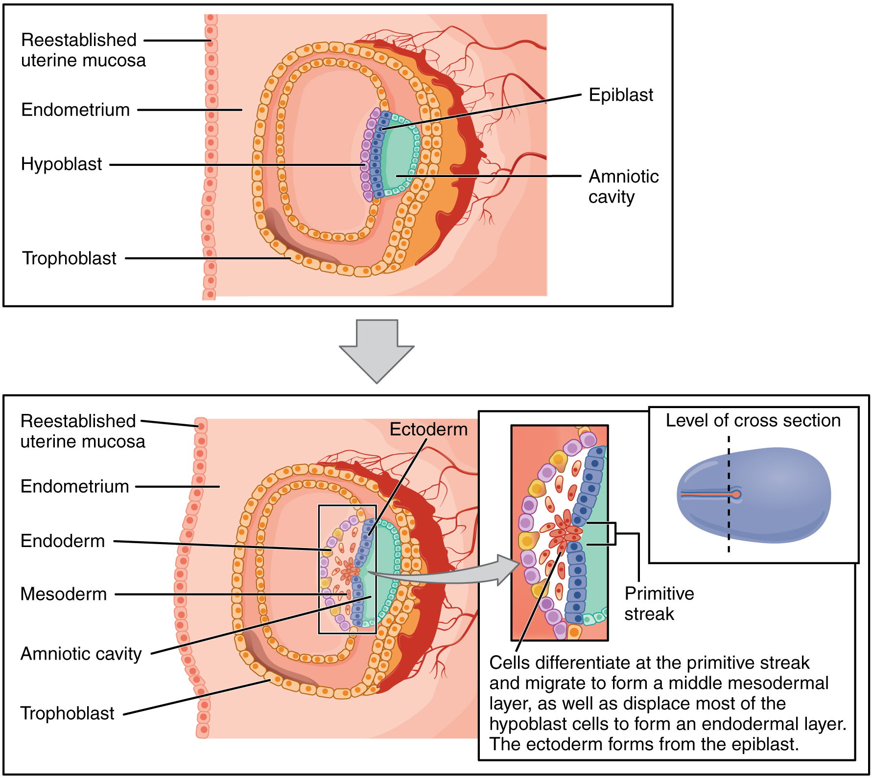
00:01 Hello! Welcome to pediatric neuropathology. 00:05 A quick overview of embryogenesis so that you have a firm understanding as to where these pathologies are arising from. 00:12 The brain tissue begins to differentiate from the ectoderm at approximately three weeks. 00:19 Weeks three to four, we have something called neurulation in which we have formation and closure of the actual neural tube. 00:27 The anterior or the rostral neural tube, the closure is within 24 days, approximately one month. 00:33 The failure of which may result in anencephaly or encephalocele, wherein the rostral portion of the spine or the neural tube. 00:46 If you get into the posterior portion, we call this the caudal portion. 00:48 This requires approximately 27 days. 00:52 So this would be a little bit longer than the anterior and, of course, failure of closure here, then puts you into the category of spina bifidas, either the occulta, the meningocele, or the myelomeningocele, which then represents both the spinal cord and the meninges, which are then protruding out and by doing so, the spinal cord being pulled out may then cause decreased ability to control one’s bladder. 01:20 Let’s go into weeks five and six. 01:22 We have vesicle formation. 01:24 By vesicle formation, prosencephalon, telenecephalon and diencephalon. 01:29 Mesencephalon remains undivided at five to six weeks. 01:34 The rhombencephalon, metencephalon, the myelecephalon is what it divides into. 01:39 And then we have holoprosencephaly, which means failure of the prosencephalon to then divide or to cleave. 01:49 Now, when you say holoprosencephaly, you should be thinking about conditions such as trisomy 13, which is your Patau or maybe fetal alcohol syndrome where you do not divide your prosencephalon into telenecephalon and diencephalon, weeks five and six. 02:07 These are clinically important. 02:10 When you get into weeks eight and thirty-two, this is cellular proliferation and migration. 02:15 This is when the sulci, which means what? The actual, the crevice or the cavity between the gyri. 02:21 They gyri means the tissue will be forming. 02:24 You have a condition called lissencephaly. 02:26 Lissencephaly means smooth cortical surface due to poor migration. 02:34 Once again, remember, I want you to think about the outer aspect of the brain, the cortex, you should have the inner crevices or the sulci, and then you have the gyri, right? But what if you don’t have the crevices and you don’t have the partitions between the gyri, and we call this a smooth cortical surface, which – that’s the pathology. 02:56 And we call the lissencephaly. 02:58 Weeks eight and thirty-two is where we are. 03:00 We have something called pachygyria, which is a large gyri. 03:05 Microgyria, smaller gyri. 03:07 And then we have schizencephaly, which is cracked brain, cleft due to defective morphogenesis. 03:15 Stroke is often responsible for this. 03:17 So think of this as being, well, you know what schizophrenia is, which is broken mentality or cracked mentality, think of this as being literally a cracked brain. 03:27 Schizencephaly. 03:29 Fascinating, isn’t it?
About the Lecture
The lecture Embryogenesis by Carlo Raj, MD is from the course Pediatric Neuropathology.
Included Quiz Questions
Neurulation takes place in which of the following time periods?
- Three to four weeks
- First week
- Early in the second trimester
- Late in the second trimester
- Third trimester
Which of the following results from the failure of the rostral end of the neural tube to close during embryonic life?
- Anencephaly
- Spina bifida
- Spina bifida occulta
- Meningocele
- Myelomeningocele
The failure of prosencephalon to develop into two hemispheres is known as...
- ...holoprosencephaly.
- ...anencephaly.
- ...telencephalon.
- ...diencephalon.
- ...myelomeningocele.
Author of lecture Embryogenesis

Carlo Raj, MD
Customer reviews
5,0 of 5 stars
| 5 Stars |
|
5 |
| 4 Stars |
|
0 |
| 3 Stars |
|
0 |
| 2 Stars |
|
0 |
| 1 Star |
|
0 |
