Playlist
Show Playlist
Hide Playlist
Landmarks and Boundaries of the Pelvis

-
Slides Landmarks and Boundaries of the Pelvis.pdf
-
Download Lecture Overview
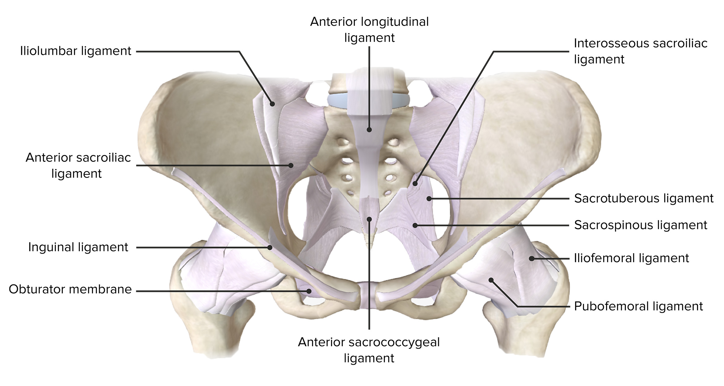
00:01 In this video, we're going to look at the pelvis. 00:04 Its various bony structures that make up the pelvis, and the boundaries of the pelvic cavity. 00:10 So let's start off by looking at the location of the pelvis, which is situated inferiorly within this abdominal pelvic cavity. 00:19 And we can see that on the screen at the moment, which is positioned anterior to the vertebral column. 00:25 So unlike the separation between the thorax and the abdomen, which has the diaphragm. 00:30 There isn't a physical structure that separates out the abdomen from the pelvis. It really is a continuous structure. 00:37 We do have some boundaries, though, that help us to recognize various regions within this combined space. 00:45 And here we can see the pelvic inlet. 00:47 The pelvic inlet here really separates the pelvic cavity from the abdominal cavity. 00:53 And here we can see parts of that pelvic cavity that extends up into the abdomen is actually part of that abdominal cavity. 01:01 It's not bound by the bony structures of the pelvis. 01:05 And this is why we call it the greater or the false pelvis. 01:08 We can see how the anterior aspect of it is much open and it can expand as it does with pregnancy, for example. 01:15 We can then see inferior and slightly posterior to the pelvic inlet. 01:19 We have the lesser or the true pelvis. 01:22 And this is part of the pelvic cavity, and it's completely bound by those various bony structures. 01:29 So here we can see the pelvic cavity, and we can look at the various boundaries. 01:34 So here again, we can introduce the pelvic inlet. 01:37 And we can then also as we have an inlet have the pelvic outlet. 01:41 So these two regions are really important, as we have the continuation of the gut tube that passes all the way down via the transverse, descending, sigmoid, parts of the colon. 01:52 All the way down and through the rectum to exit the pelvis via the anus at the pelvic outlet. 01:58 So there needs to be this continuous space that allows foodstuffs, digested foodstuffs, to leave the body cavity. 02:05 It's also important for reproductive and urinary function that we have this continuation from the abdomen down through the pelvis and then to exit the body. 02:15 We can see laterally within this space. 02:16 We have pelvic walls. And we can see these if we have a look at the superior view of the pelvis. 02:23 Posteriorly, we can see we have S1. So, the vertebral body of S1. 02:28 Lateral to that on both sides, we have the Ala of the sacrum, and that then runs anteriorly all the way towards the pubic crest via the pelvic brim, highlighted here in red. 02:39 Then most anteriorly, uniting two halves of the pelvis, we have the pubic symphysis. 02:45 So, from posterior to anterior, we have the vertebral body of S1, remember, or the sacral vertebrae are fused to form the sacrum. 02:53 We have the Ala of the sacrum. 02:54 We have the pelvic brim, pubic crest, and then the pubic symphysis. 02:59 If we then have a look at this in section, we can see where the pubic body has been section. 03:04 So, this is where the pubic symphysis would attach each sides of the pelvis. 03:09 Then moving laterally, we have the superior pubic ramus. 03:13 Moving inferiorly, we have the inferior pubic ramus. 03:16 And if you've looked at the lectures on the low limb, these terms should be familiar with you. 03:22 We then have the continuation of the inferior pubic ramus as it goes all the way around to form the ishiopubic ramus And these bony structures then form the obturator foramen. 03:32 An important foramen that allows structures to pass from the pelvis all the way down into the lower limb like the obturator nerve. 03:40 If we then continue looking at these bony structures, most superiorly we have the greater sciatic notch, and then below that we have the lesser sciatic notch. 03:49 These two notches are important as they allow structures to pass between the gluteal region of the lower limb and the pelvis, and also of the pelvis and a structure, a space, we'll see later on, the perineum. 04:02 So, these are really spaces that allow structures to pass between the pelvis and related areas. 04:08 We have the greater sciatic notch and the lesser sciatic notch separated by the ischial spine. 04:13 We then have most inferiorly, the ischial tuberosity. 04:17 Posteriorly, we have the sacrum, and then connecting these bony structures to the sacrum, we have a series of ligaments. 04:24 So, from the ischial spine to the sacrum, we see highlighted here the sacrospinous ligament. 04:29 And then from the ischial tuberosity to the sacrum, we have the sacrotuberous ligament. 04:35 And these are now converting those notches into foramen. 04:39 So, here we have the greater sciatic foramen, and here we have the lesser sciatic foramen. 04:44 We'll come across these spaces in more detail as we progress through these pelvic videos. 04:50 Now, let's have a look at the pelvic outlet. 04:52 So this is a space that allows structures to really exit the pelvis and either enter into the perineum or go into the outside world. 04:59 Here we can see the pelvic outlet. 05:01 Anteriorly is demarcated by the pubic symphysis. 05:04 We then have the ischiopubic remi on either side. 05:08 We then have the ischial tuberosity. 05:10 And then that ligament that connected the ischial tuberosity to the sacrum, we have the sacrotuberous ligament. 05:16 With most posteriorly, we have the coccyx. 05:18 So here we can see the boundaries that form the pelvic outlet. 05:23 So, now let's talk about the pelvic floor. 05:26 We'll talk about this in a lot more detail in a later lecture. 05:29 But really, this is a muscular fibrous layer that sits over the pelvic outlet, and it serves to hold the pelvic organs in place. 05:39 We can see here this thin muscular layer is stretching from the lateral aspect of the pelvis from each side, and they unite in the midline. 05:48 And later on, we'll hear about structures like the perineal body, which is the central fibrous structure that helps to unite these two muscular layers in the midline. 05:58 But importantly, the pelvic floor has a couple of apertures that allows various structures to pass through them from either the pelvis into the perineum or from the pelvis into the outside world. 06:08 But we'll cover the pelvic floor in more detail later on. 06:11 For the time being, just remember, it's a muscular layer that helps to support the pelvic structures in place. 06:19 Sitting superior to the pelvic floor is the pelvic cavity as we've just demarcated with its lateral anterior and posterior boundaries. 06:26 And within the pelvic cavity, we'll find organs in relation to digestion, with the rectum and the anus with reproduction. 06:34 Be that the uterus, be that the vagina, or the prostate in the male, and also to do with maturation with the bladder. 06:42 So the pelvis is an important structure, which we'll talk through in the next few slides. 06:48 Sitting underneath the pelvic cavity separated by the pelvic floor is the perineum. 06:54 And the perineum is an important structure for allowing those structures to pass from the pelvis to the outside world. 07:01 And as an important function with both reproduction, micturition and with the removal of digested foodstuffs. 07:07 We'll come back to the perineum later on.
About the Lecture
The lecture Landmarks and Boundaries of the Pelvis by James Pickering, PhD is from the course Bony Pelvis and Pelvic Floor.
Included Quiz Questions
What is the most anterior part of the pelvis?
- Pubic crest
- Pelvic brim
- Ala of sacrum
- S1 body
- L1 body
What separates the greater and lesser sciatic notches?
- Ischial spine
- Ischial tuberosity
- Superior pubic ramus
- Pubic body
- Inferior pubic ramus
What is the location of the perineum with respect to the pelvic floor?
- Inferior
- Superior
- Lateral
- Medial
- Oblique
Author of lecture Landmarks and Boundaries of the Pelvis

James Pickering, PhD
Customer reviews
5,0 of 5 stars
| 5 Stars |
|
5 |
| 4 Stars |
|
0 |
| 3 Stars |
|
0 |
| 2 Stars |
|
0 |
| 1 Star |
|
0 |
