Playlist
Show Playlist
Hide Playlist
Hip Joint

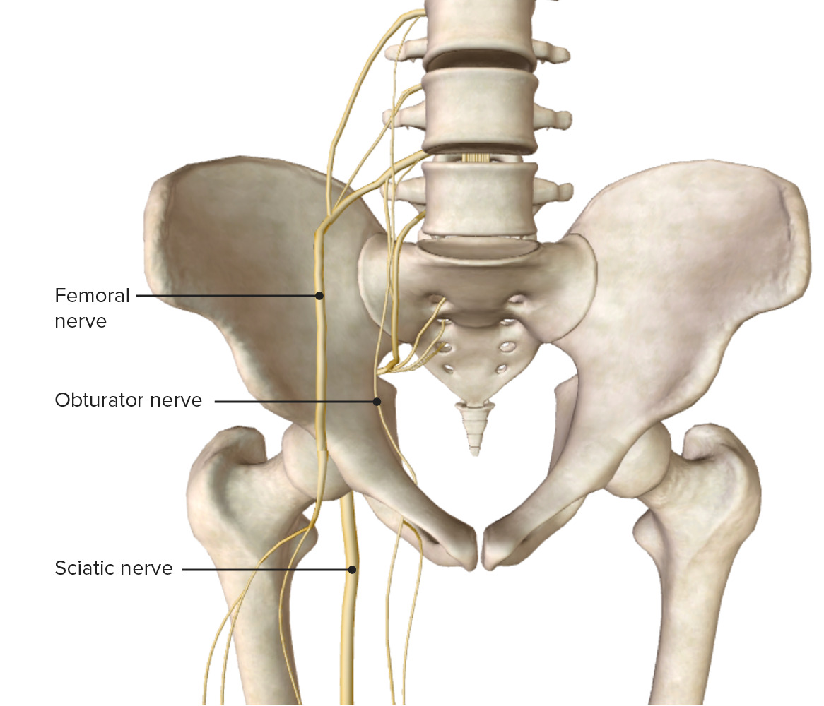
00:01 Let's start with the Hip joint. 00:04 So the Hip joint is a synovial ball and socket joint. 00:09 It's the articulation between the head of the femur into the acetabulum of the pelvis bone. 00:16 So here we can see as we separate the femur from the pelvic bone, we can see we have the head of the femur, and here we have the acetabulum. 00:24 If we look at the lateral view, we can see the acetabulum here indicated with this black dotted line. 00:31 To the right hand side of the screen, we have the anterior aspect where we can see the pubic symphysis just protruding out. 00:38 And the ischial tuberosity is down in the lower left hand side projecting posteriorly. 00:43 But here we have the acetabulum. 00:45 This circle indicated via the black dotted line. 00:49 It's made up of those three bones that we've spoken about that form the pelvic bone. 00:54 We have the ilium in green, the ischium in blue, and the pubis bone here in red. 01:00 These three surfaces give rise to what is the articular surface of the hip joint, which is this lunate surface, that has this very characteristic shape. 01:10 We also have the acetabular fossa. 01:13 And this doesn't actually have any articulation with the head of the femur. 01:17 Surrounding these surfaces is the hyaline cartilage. 01:21 And that creates that smooth surface for the head of the femur to articulate with. 01:26 Running circumferentially around the acetabulum, we have the acetabular labrum and this increases the articular surface. 01:34 Increasing the bone congruency, that space in which the head of the femur can articulate. 01:40 It helps to further stabilize this important joint. 01:44 Just connecting that lunate surface where we've got the hyaline cartilage, we have an important ligament. 01:50 This is the transverse acetabular ligament, and it's positioned posteriorly where there is a deficiency in this hyaline cartilage. 01:59 In there, we can then see the acetabular foramen, and we'll come back to this in later detail. 02:04 Running through from this acetabular fossa, we have the ligament of the head of the femur. 02:09 And that passes into the fovea, which we'll see later on in the head of the femur. 02:15 So now let's have a look at the head of the femur. 02:17 And this is a right femur, we're looking at his anterior surface. 02:21 Here, we have hyaline cartilage covering the articular surface of the head of the femur. 02:27 And here we have that fovea, which is for the ligament of the head of the femur, and this helps to hold the femur in position. 02:35 Here we can now see the ligament of the head of the femur on the screen. 02:39 Here we can see the head of the femur is positioned with in the acetabulum. 02:44 And surrounding it, we have a joint capsule. 02:47 So this is a joint capsule that surrounds that joint and helps to hold the head of the femur in position. 02:54 We can see it's attaching medially on to the pelvic attachment around the margin of this acetabulum. 03:00 And the adjacent margin occurs around the obturator foramen. 03:04 We also have it attaching onto the intertrochanteric line anteriorly on the femur, and also on the neck of the femur posteriorly. 03:14 Now, let's turn our attention to the ligaments that help to support the hip joint. 03:19 These ligaments are very much named after the bones which they originate from. 03:24 So here we have the Pubofemoral ligament. 03:27 Here we have the Iliofemoral ligament. 03:29 And more posteriorly, we have the Ishiofemoral ligament. 03:33 So these ligaments are running alongside the joint capsule. 03:36 We have three of them, which originate from those three bones that form the acetabulum. 03:41 Iliofemoral, Pubofemoral, and Ishiofemoral ligaments. 03:46 Here we can see the blood supply that goes to supply the joint capsule around the hip. 03:50 It's originating from numerous branches including the obturator artery, the femoral artery, and the deep artery of the thigh. 03:58 All of these main trunks give rise to numerous branches that surround the hip joint and give it a wide range of blood supply. 04:06 For example, we have the superior and inferior gluteal arteries originating from the internal Iliac artery. 04:13 We then have the two circumflex arteries, medial and lateral, which are coming from the deep artery of the thigh. 04:19 So we spoke previously in the overview of the blood supply to the lower limb that these form an important and that's demotic loop around the hip joint. 04:30 Specifically, we touched on how the obturator artery passing through the obturator canal that deficit in obturator membrane. 04:37 How its two anterior and posterior branches form an important anastomosis. 04:42 This gives rise to an acetabulum branch that passes all the way to the head of the femur running within the ligament of the head of the femur. 04:51 So an alternative blood supply that's running straight in to the substance of the femur itself. 04:58 So now, let's look at the movements that occur at the hip joint. 05:02 We have the femur moving anteriorly here within the sagittal plane, and that is going to be Flexion. 05:08 We also have the femur moving posteriorly here within the sagittal plane, and that is going to be Extension. 05:14 We also have the femur able to move towards the midline and we call this Adduction. 05:19 We also have the femur then moving laterally away from the midline, and this is going to be Abduction. 05:27 We also have a degree of rotation at the hip joints, we have external or lateral rotation, and we can also have medial or internal rotation. 05:36 So a range of movements that can occur at the hip joint.
About the Lecture
The lecture Hip Joint by James Pickering, PhD is from the course Joints of the Lower Limbs.
Included Quiz Questions
What is one difference between the lunate surface and the acetabular fossa of the hip joint?
- The lunate surface is an articular surface.
- The lunate surface is round.
- The lunate surface is cartilaginous.
- The lunate surface is fixed.
- The lunate surface is composed of hyaline cartilage.
What are the ligaments of the hip joint? Select all that apply.
- Iliofemoral ligament
- Pubofemoral ligament
- Ischiofemoral ligament
- Ischiopubic ligament
- Femoroacetabular ligament
Which artery passes through the ligament of the head of the femur?
- The acetabular branch of the obturator artery
- The acetabular branch of the superior gluteal artery
- The acetabular branch of the femoral artery
- Medial circumflex artery
- Lateral circumflex artery
Author of lecture Hip Joint

James Pickering, PhD
Customer reviews
5,0 of 5 stars
| 5 Stars |
|
5 |
| 4 Stars |
|
0 |
| 3 Stars |
|
0 |
| 2 Stars |
|
0 |
| 1 Star |
|
0 |
