Playlist
Show Playlist
Hide Playlist
Arteries and Veins: Structure and Characteristics

-
Slides Structure-Function Relationships Cardiovascular System.pdf
-
Download Lecture Overview
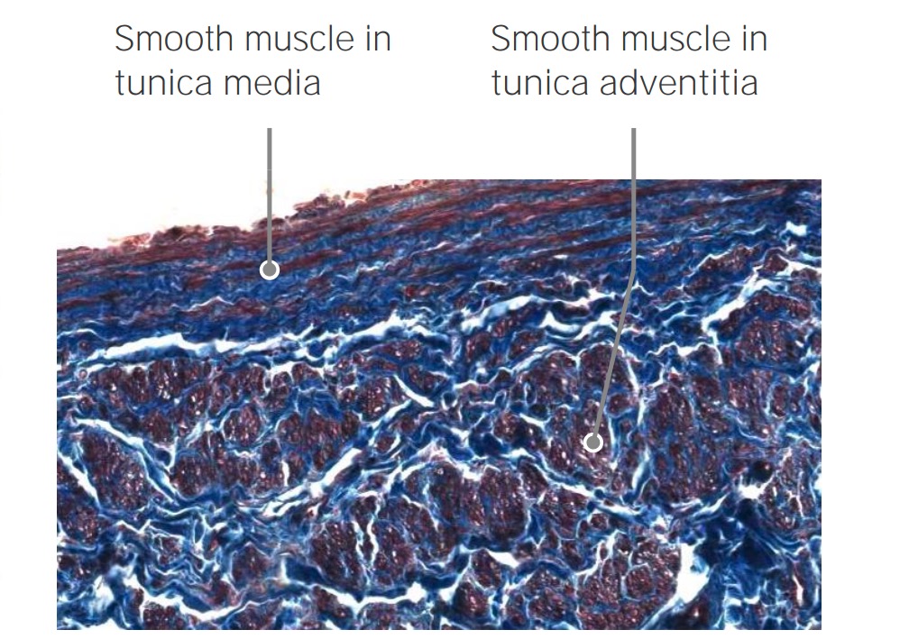
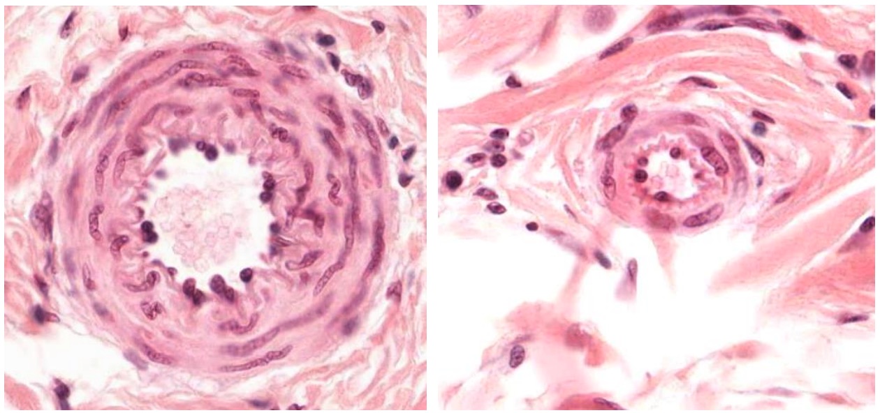
00:01 Hello and welcome. 00:02 We're going to talk about something that is very near and dear to my heart, which is the cardiovascular system. 00:08 And hopefully it's near and dear to your hearts as well. 00:12 To really understand the pathology of heart and vessel, we really have to understand the structure and function relationships. 00:19 And actually, the cardiovascular system is beautiful in that respect. 00:23 Because if you were going to design the system, this is the way you would do it. 00:26 It's very logical, and it's very physiologic, and it works terrific. 00:32 And it will last most of us 80-90, hopefully, 100 years. 00:36 With that preface, let's get going. 00:38 Here's the kind of general framework roadmap for where we're going today. 00:43 We're going to do an organization of the vasculature so understand how vessels are assembled into a closed circuit. 00:49 We'll talk about the vessel lining endothelium, which is actually a whole lot more important than just a vessel lining. 00:56 And then we'll talk about basic structures of the heart. 00:59 First up is the organization of the vasculature. 01:02 We are seeing here a fairly complex picture, with the beating heart centrally as it should be. 01:08 Blood comes in from the superior and inferior vena cava bringing blood back from the entire body into what's going to be the right atrium, the right ventricle, and eventually it gets pumped out to the lungs, and then eventually returns from the lungs to the left ventricle, and out the aorta. 01:28 Pulmonary artery is leaving from the right ventricle into the lungs, and it's still carrying deoxygenated blood. 01:35 An important point in the red box at the bottom, arteries are not always oxygenated, and veins are not always deoxygenated. 01:44 So for example, when we look at the pulmonary artery, it is a deoxygenated artery. 01:49 Mainly, we talk about arteries is taking blood away from the heart and veins bringing blood back to the heart. 01:56 So again, not always just oxygenated and deoxygenated. 02:01 The aortic arch, the aorta is bringing blood now out of the left ventricle, squeezed at 120 millimeters of mercury, and going out and perfusing the entire body. 02:13 Going out through the descending aorta, and then into the lower organs, but also perfusing head and neck and the vessels of the upper extremities. 02:25 An important kind of general conceptual thing: Veins are low pressure. 02:30 They have very high capacitance, about two thirds the total blood volume lives in the veins at any particular moment in time. 02:39 On the other hand, the arterial system is high pressure. 02:43 It has to transmit blood to the entire body at a pressure sufficient to move it through tiny capillaries. 02:50 So it's a high pressure system but low volume. 02:54 Okay, let's get down into some of the nitty gritty. 02:58 When we're talking about the high pressure system, the arterial side, it's got a certain organization. 03:04 And we're also going to talk about the venous side, and it has a certain organization. 03:09 They have the same general three layers, but they are put together slightly differently. 03:15 Let's look now at the left hand side at an artery, high pressure system. 03:19 Vessels everywhere in the body are lined by an endothelium, a simple squamous epithelial layer. 03:27 It sits on top of a basement membrane, or a basal membrane, or a basal lamina. 03:33 They're all synonymous. 03:36 That is then identified the endothelium. 03:40 And the basement membrane that it's sitting on are called the intima. 03:44 This is the innermost layer of all blood vessels, the intima. 03:49 Demarcating in the arterial side of the circulation between the intima and the next layers is going to be an internal elastic membrane, or an internal elastic lamina. 04:02 That's going to have a little bit of elastic structure to it, and be able to expand, and contract, and recoil. 04:10 That demarcates the intima from the media. 04:13 The media's just like it sounds, it's the middle layer. 04:17 It's composed of lots, and lots, and lots of smooth muscle cells arranged in a circumferential organization. 04:26 Demarcating the media from the most external layer is going to be the external elastic lamina or external elastic membrane. 04:34 This is quite variable. 04:35 It may not be well defined in many arteries, but there's going to be elastic tissue in that location. 04:42 And then outside of that elastic membrane is going to be the adventitia. 04:48 The adventitia is going to be loose connective tissue, and also have additional structures including nerve, connective tissue, and vasa vasorum vessels. 04:58 We'll talk more about that shortly. 05:00 That's the artery, high pressure, low volume. 05:05 So, the intima, let's get a little bit more intimate with the intima. 05:09 It's specialized lining for all blood vessels. 05:14 The gene expression profile. 05:16 The behavior, the appearance varies very much in the intima depending on where you are in the vascular tree. 05:26 So if you are in the aorta, it can be quite different than if you are in the capillaries. 05:33 So the endothelial expression, the behavior, everything else, varies depending on where you are. 05:39 And we'll cover that in more detail in a moment. 05:42 The media. Concentric layers of smooth muscle cells that wrap around the vessel lumen. 05:48 And this is going to allow us to impart contraction, or relaxation, That will allow us to have higher pressure or lower pressure. 05:59 There's a very high elastic content, also within many vessels, aortic vessels, and arterial vessels. 06:07 And that allows us to have recoil. 06:09 We'll talk in a moment about how the heart is now pulsatile? And it goes from a higher pressure to a lower pressure from systole to diastole. 06:18 And when it goes through systole, the vessels expand ever so slightly. 06:22 And then we go to diastole, they contract because of the elastic content that allows us to propel blood forward. 06:32 There's a lot of input from the autonomic nervous system in the arterial circulation. 06:37 This is how we control blood pressure. 06:39 We want to ensure that we get blood to every last little nook and cranny of the body. 06:44 And we need to regulate at a local level and also a systemic level, how tight or what kind of tone the vessels have? Are they squeezed? So they have a high tone. 06:55 Are they relaxed, so they have a low tone? Out to the adventitia. 07:03 So this contains the nerve fibers and the vessels of the vasa vasorum. 07:07 Literally, vasa vasorum means the vessels of the vessel. 07:12 Some arteries are so thick-walled that they need to have their own blood supply. 07:18 They cannot depend on diffusion, just from the lumen. 07:22 So, we have vessels of the vessel. 07:25 These have vasa vasorum that will perfuse those medial smooth muscle cells. 07:32 Again, as already been mentioned, we may separate the media from the adventitia by an external elastic lamina, that may not always be the case. 07:45 Okay, on the left hand side, that's the artery. 07:48 On the right hand side, let's dive a little bit deeper into the venous structure. 07:53 Veins have the same structure, just different amounts of the various components. 08:00 So the endothelium got to be there. And that's a truism. 08:03 If you don't have an intact endothelium you don't have a happy blood vessel. 08:09 So there's always going to be endothelium lining every part of the cardiovascular system. 08:15 There will be the endothelium is an epithelium and it sits on a basement membrane or basal membrane. 08:22 And then there's going to be varying amounts of a media. 08:25 Note: It's much thinner, and it's not going to be as well organized as the media that we saw in the arterial circulation. 08:33 There will also be an adventitia. 08:36 Connective tissue that allows us to kind of organize the vessel into a tube. 08:41 Alright, so same general structures, but there's different amounts of the various components. 08:51 What does this look like in real life? In the lower left hand corner is an artery, cut in cross section. 08:57 And we can see even from here that it looks more well-defined in terms of the three layers versus the vein which is going to be in the upper right hand corner. 09:09 We're going to look at each one of those in turn. 09:10 So this is a low power view. 09:12 This is an artery and a vein sitting in fat. 09:15 And we're gonna be looking now at the endothelial cell layer. 09:18 Again, endothelium very thin, simple squamous, epithelium, sitting on a basal lamina. 09:25 We have the Internal Elastic Membrane that demarcates the intima from the media. 09:31 We then have the smooth muscle media the various layers are called tunicus so you can have the tunica intima, the tunica media, the tunica adventitia. 09:42 Where you can just say layers, unless you like Latin. 09:46 And then you have the External Elastic Membrane and finally, we have the adventitia. 09:50 So we have all the various layers here well demarcated. 09:55 Note: When we look at that artery, you can see the vessel lumen size. 09:59 Compared to the vein, it is much smaller. 10:03 This is going to be a higher pressure system. 10:06 We need to kind of reinforce the layers of the vessel, because it's higher pressure. 10:11 And that's why there's more smooth muscle. 10:14 That's why there's more of a connective tissue adventitia. 10:18 Now, if we go up to the vein, there— bigger lumen, higher capacitance, greater volume, lower pressure. 10:26 So the wall doesn't have to be as robust. 10:28 It doesn't have to be as thick. 10:31 Still has an endothelium, single cell layer, same deal, sitting on a basal lamina. 10:36 And then that's it. That's the intima. 10:39 Now, we're into the smooth muscle media, the tunica media, that layer not as well organized, not as much as in the artery because it doesn't have to maintain the same pressures. 10:51 It doesn't have to. 10:52 It has to actually be more expansile, more capacitance to hold greater amounts of blood. 10:58 And then we're into the adventitia. 11:02 Also shown here is the nerve. And again, vessels are innervated. 11:07 That's how we can control to some extent, the arterial smooth muscle tone, but also venous tone. 11:16 There are smooth muscle cells there. 11:18 Vein cannot squeeze as hard as an artery can, but it can still modulate how well it squeezes and nervous input will influence the tonicity the tension of how much the smooth muscle contracts in either vessel.
About the Lecture
The lecture Arteries and Veins: Structure and Characteristics by Richard Mitchell, MD, PhD is from the course Structure-Function Relationships in the Cardiovascular System.
Included Quiz Questions
What is the name of the innermost layer of all blood vessels?
- Tunica intima
- Tunica media
- Internal elastic membrane
- Tunica adventitia
- External elastic membrane
Which vessel layer is correctly paired with its descriptor?
- Adventitia - loose connective tissue that contains nerve fibers and the vasa vasorum
- Intima - all vessels have the same gene expression profile for this layer
- Media - the single layer of cells that is directly adjacent to blood flow
- Endothelium - circular muscle layer that aids in vasoconstriction
- External elastic membrane - the major muscular component of both veins and arteries
What is one of the major differences between arteries and veins seen on microscopy?
- The amount of tissue in each layer is greater in arteries.
- The capacitance of the arterial lumen is greater.
- Veins have two main layers, while arteries have three.
- The tunica media of arteries is not as well organized as the tunica media of veins.
How does the structure of the media contribute to the functions of the arterial and venous systems?
- It allows the arterial system to be a high-pressure system and the venous system to be a high-capacitance system.
- It allows the arterial system to be a low-pressure system and the venous system to be a low- capacitance system.
- It allows the arterial system to be a high-pressure system and the venous system to be a low- capacitance system.
- It allows the arterial system to be a low-pressure system and the venous system to be a high- capacitance system.
- It allows the arterial system to be a high-capacitance system and the venous system to be a high-pressure system.
Author of lecture Arteries and Veins: Structure and Characteristics

Richard Mitchell, MD, PhD
Customer reviews
5,0 of 5 stars
| 5 Stars |
|
1 |
| 4 Stars |
|
0 |
| 3 Stars |
|
0 |
| 2 Stars |
|
0 |
| 1 Star |
|
0 |
The lecturer did a great job explaining the anatomy of the heart, and with ease too.
