Playlist
Show Playlist
Hide Playlist
Surface Anatomy of the Lower Limbs
-
Slide Surface Anatomy Lower Limbs.pdf
-
Download Lecture Overview
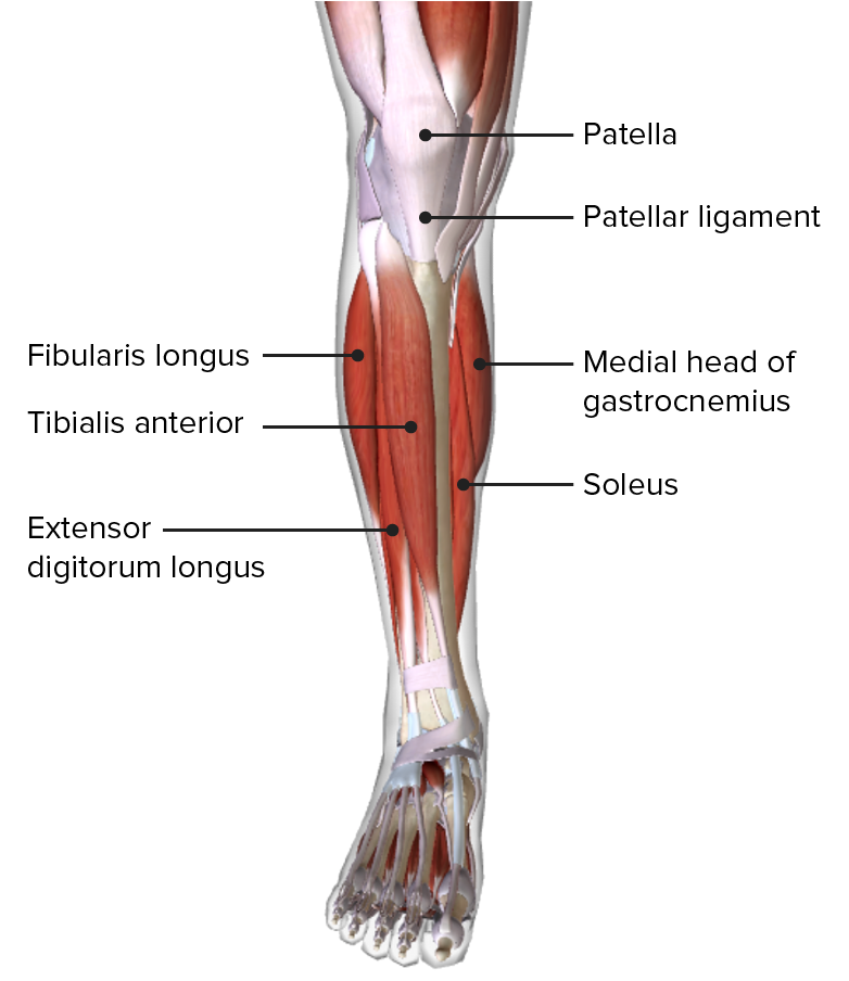
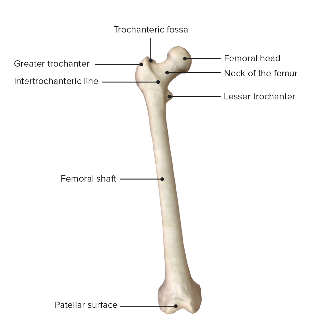
00:01 In this lecture, we're going to look at the surface anatomy of the Lower Limb. 00:06 We'll also look at the specific bony structures that make up the lower limb all the way from the hip bone down to the foot where we have the tarsals and metatarsals, etc. 00:16 So we'll look at the surface anatomy of the lower limb and then move on to look at the individual bones that make up the lower limb. 00:22 So on the screen at the moment, you can see the anterior and posterior surface of a right lower limb. 00:29 So here we can see some broad regions that we'll go into a little bit more detail over the next few slides. 00:34 But we can see that we have a thigh region, there's the knee separating the thigh from the lower aspect of the lower limb which we call the leg. 00:43 And here we can see you then have the dorsum of the foot. 00:46 So we move to the foot region, we swapped the word anterior and look at the dorsal surface. 00:51 So we can see we've got a thigh, knee, leg, and then a foot region on its anterior surface. 00:57 If we look at the posterior surface, we can see they're similar structures, similar surfaces. 01:01 So we have a thigh region, we have a lower limbs leg region, and we also have a gluteal region at the very top of the lower limb. 01:09 And then in between the thigh and the leg region. 01:12 On the posterior surface, we have a region called the popliteal region and deep within there we have the popliteal fossa. 01:19 And that's an important structure we'll talk about later on. 01:22 We then have a region known as the calcaneal region, which some of you may be familiar as the Achilles tendon area where that Achilles tendon importantly runs down from the posterior aspect of the leg onto the sole of the foot which you can see on the bottom of the screen. 01:38 So a number of kind of broad areas that make up the lower limb or thigh, knee, leg and foot region on the anterior surface, a gluteal, thigh, popliteal region, leg, and then the sole of the foot, we can see here on the posterior surface. 01:53 So let's concentrate on the anterior surface of the thigh region which is indicated by this green box. 01:59 And specifically, let's start by looking at some of the regions here and some specific structures, which we can see protruding through the surface of the skin to create these landmarks, and what we can see shaded in here in green is the tensor fasciae late muscle. 02:14 This is an important muscle as it gives rise to a very thick band of fasciae which is a tissue that spreads over the anterior surface of the thigh down the lateral aspect of the knee. 02:27 And what this does is a couple of things, it helps to hold all those muscles tightly against the femur deep within the thigh area we can see there. 02:36 But it also helps to create big stability of this region. 02:40 So it helps to keep stability of the knee region and of the hip region because obviously that's important as you're standing up, and gravity is pushing your body down. 02:48 It's important you have the stabilizers to maintain your posture. 02:52 So tensor fasciae late muscle, we'll see it again when we look at the muscles of the thigh, but it's an important landmark you can see protruding through the surface of the skin there. 03:01 Another muscle we can see which is making an impact on the skin is sartorius muscle. 03:06 This is running in furrow medially down from the surface of the thigh we can see there. 03:14 We can see another series of muscles, rectus femoris, vastus lateralis muscle, vastus medialis muscle and quadrices femoris tendon. 03:23 These are a whole series of important structures which will spend more time going through as we move through the muscles and the various tendons of the thigh region in later lectures. 03:34 If we now move inferior to the knee joints, we can see we have the quadrices femoris tendon. 03:40 This is that tendon that's formed by those four muscles that pass over the patella and then insert into the tibia. 03:47 We can see the patella here and there is that patellar ligament. 03:50 This is one continuous tendon that passes through the patella, the patella then ossifies during development to form that small bone we have on the anterior aspect of the knee. 04:01 And that tendon then continues down onto the tibial tuberosity. 04:05 We have a series of muscles which we can see on the anterior surface of the leg here. 04:09 We have tibialis anterior muscle, we have the anterior border of the tibia we can see here. 04:15 We have the lateral and the medial malleoli, we can see both of the inferior aspect of the leg. 04:22 Now let's spin the leg around, we're still looking at a right lower limb but this time we're looking at its posterior surface. 04:29 And again within this green box, we can see some structures indicated. 04:34 We have a very large muscle which is really forming the buttock region most superiorly within the thigh and that is the big muscle of gluteus Maximus. 04:44 We have a series of gluteal muscles and this is the most superficial, it's also the largest is the gluteus Maximus muscle. 04:52 Some other features we can do estimate may carry we have the greater trochanter of the femur. 04:56 We then have a cleft of the gluteal muscle as it then extends down into the hamstring portion of the thigh. 05:03 Here we have the iliotibial tract which is associated with the tensor fasciae late muscle I mentioned previously here we can observe it on the posterior aspect. 05:12 And then if we extend down onto the posterior surface of the leg, we can see some very prominent muscles here, especially in athletes that do considerable amount of running, we can see the popliteal fossa region most superiorly and then we have these big two heads of gastrocnemius. 05:30 The single muscle has two heads, the medial and lateral head and you can see gastrocnemius there. 05:36 Gastrocnemius along with some other muscles we'll come to later on gives rise to the Achilles tendon, or the calcaneal tendon and that attaches to the calcaneal tuberosity, or the heel bone that some of you may be familiar with. 05:49 So that's a brief overview of the various surface landmarks that one can see when they're looking at the anterior and posterior region of the lower limb. 06:00 So now let's have a look at the bones that make up the lower limb. 06:03 And there's, as you can imagine, similar to the upper limb, there are a considerable number of bones here that you need to know about. 06:11 And it's important, you should look at your own curricula, your own learning objectives to specifically know which structures you need to know about because there's lots of bony structures, bony little protuberances on each of these bones, and we'll go through them but please make sure you know what you do need to know as part of your course. 06:28 So let's start having a look at the bones of the lower limb we can see here, and we start most superiorly with the pelvic or the hip bone. 06:36 And obviously we have two of these that make up that whole pelvis. 06:40 We then extend inferiorly down to a very large six strong bone which is the femur. 06:46 We then have the patella which we spoke about previously. 06:50 Then we have more medially positioned within the leg, we have the tibia, and laterally we have the much thinner, much smaller fibula, which is really there to help increase the surface area for muscle attachments. 07:02 We then have a whole series of foot bones, the metatarsals, the tarsals and the individual phalanges that make up the foot we can see those later on. 07:12 But we have a whole series of foot bones that the most distal position within the lower limb.
About the Lecture
The lecture Surface Anatomy of the Lower Limbs by James Pickering, PhD is from the course Osteology and Surface Anatomy of the Lower Limbs.
Included Quiz Questions
What is the name of the posterior surface region of the knee?
- Popliteal region
- Gluteal region
- Posterior thigh region
- Posterior leg region
- Calcaneal region
What is closely associated with the tensor fasciae lata muscle?
- Iliotibial tract
- Gluteal fold
- Popliteal fossa
- Medial head of the gastrocneumius
- Achilles tendon
Which bone is most inferior?
- Tibia
- Patella
- Femur
- Pelvic bone
- Sacrum
Customer reviews
5,0 of 5 stars
| 5 Stars |
|
5 |
| 4 Stars |
|
0 |
| 3 Stars |
|
0 |
| 2 Stars |
|
0 |
| 1 Star |
|
0 |


