Playlist
Show Playlist
Hide Playlist
Arteries of the Gastrointestinal Tract

-
Slides Arteries of the Gastrointestinal Tract.pdf
-
Download Lecture Overview
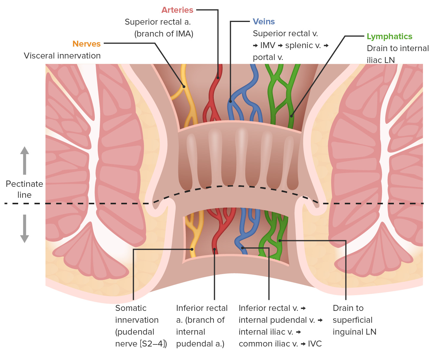
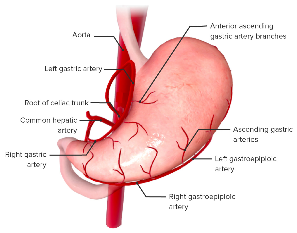
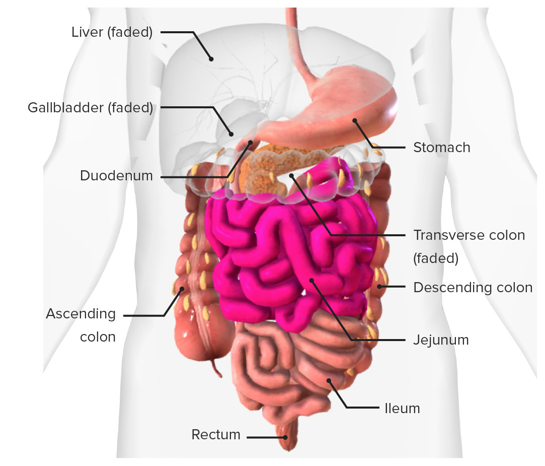
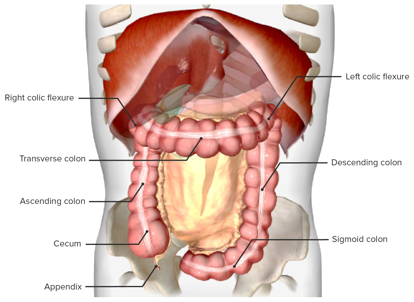
00:01 So now let's turn our attention to the blood supply and specifically the arterial supply of the entire gastrointestinal tract. 00:11 So previously, we've looked at the blood supply to various specific organs. 00:15 And we've touched upon the distribution of the arteries to the foregut, the midgut, and the hindgut. 00:22 Now, let's bring all that together and look at it as one piece of information. 00:27 So on the screen at the moment, we can see at the bottom of the screen, the abdominal aorta. 00:32 At the top of the screen, we can see sitting anteriorly, to the abdominal aorta, the entire gastro intestinal tract. 00:41 And what we've done is we've represented it as one long tube. 00:45 But we can split up this gastrointestinal tract into to the foregut, midgut, and hindgutt. 00:53 Now, each of these areas, each of these portions of the gastrointestinal tract are going to be supplied by a specific unpaired branch of the abdominal aorta. 01:06 That means that each one of these areas is supplied by a single branch coming from the aorta. 01:13 So at the bottom of the screen, we can see the abdominal aorta has passed through the diaphragm through their aortic hiatus at around about T12. 01:23 We can see that coming off just as it passes through the aortic hiatus within the diaphragm, it gives rise to the celiac trunk. 01:32 The celiac trunk is going to be the main unpaired blood vessel that supplies all of the foregut. 01:39 Coming off the abdominal aorta, slightly more inferiorly, at approximately the level of the first lumbar vertebrae, we have the superior mesenteric artery. 01:49 This blood vessel is going to supply the entirety of the midgut. 01:54 We then finish with the hindguts. 01:56 And coming off the third lumbar vertebral level of the aorta, we have the inferior mesenteric artery. 02:04 So these three unpaired blood vessels go and supply the foregut, the midgut, and hindgut with the celiac trunk, the superior mesenteric and the inferior mesenteric arteries supplying those regions. 02:18 As we've seen, and what you've seen in previous lectures, is there are these things called anastomosis. 02:23 And this is where interconnecting branches from two different sources joined together and how to add some connectivity to this blood supply. 02:34 They help to rely for some redundancy in the system. 02:37 So if there's a potential blockage somewhere, there's a secondary pathway that this blood can pass to that location. 02:44 So here we have the foregut, midgut, and hindgut with some anastomosis between those two. 02:50 And we've seen those anastomoses occur around the major duodenal papilla, and around the transverse colon, 2/3 and 1/3. 02:59 They're the separation points between foregut and midgut, midgut and hindgut, and the blood supply to those areas are therefore coming from these blood vessels. 03:10 Bridging across those two different regions we have, as I said, various anastomosis. 03:17 Let's have a look at this in some detail. 03:19 Let's have a look at the actual anatomy and how it supplies these regions. 03:23 So here we can see we have the abdominal aorta, passing through the aortic hiatus at the level of the 12 thoracic vertebra. 03:31 Here we can see at the fourth lumbar vertebrae, and just at the intervertebral disc between the fourth and fifth, we have the abdominal aorta, bifurcating into the two common ilac arteries. 03:43 They go on to supply the pelvic organs and the lower limb. 03:47 We'll talk about those later. 03:50 Let's have a look then, at the celiac trunk supplying the foregut. 03:54 And which structures reside within the foregut? We have the abdominal esophagus, we have the liver, we have the stomach. 04:02 We also have the spleen, we have the pancreas, we have the gallbladder, and we have the first couple of bits of the duodenum. 04:10 We have the first part and the first half of the second part up to the major duodenal papilla. 04:19 And here we can see that landmark, the major duodenal papilla, separating the foregut from the midgut. 04:27 Now, let's have a look at the midgut and what structures reside within this area. 04:32 So, the midgut is running very much from that major duodenal papilla. 04:36 But now inferior to it. 04:39 So the second bit of the second part, and then the third and fourth parts of the duodenum. 04:45 They're part of the midgut. 04:47 We also then have all of the small intestines. 04:50 We've got the jejunum and ileum. 04:52 And then the appendix, the cecum, ascending colon, and then up to the first two-thirds of the transverse colon. 05:01 So that part of the transverse colon that's coming from the hepatic flexure, and then working all the way to the splenic flexure, two-thirds of that distance is classically the midgut. 05:12 Let's then turn to the hindgut and which structures reside within this area. 05:18 So we finished on the midgut with the first two-thirds of the transverse mesocolon. 05:25 Now we have the distal third of the transverse colon. 05:28 And this is the beginning of the hindgut and supplied by the inferior mesenteric artery. 05:34 We then have the descending colon, we have the sigmoid colon, and then we have the rectum and the upper part of the anal canal.
About the Lecture
The lecture Arteries of the Gastrointestinal Tract by James Pickering, PhD is from the course Vascular Supply of the Abdomen.
Included Quiz Questions
At which vertebral level does the descending aorta pass through the diaphragm?
- T12
- T6
- T8
- T10
What is the principal artery of the foregut?
- Celiac trunk
- Superior mesenteric artery
- Inferior mesenteric artery
- Splenic artery
- Common hepatic artery
What is the vertebral level where the inferior mesenteric artery originates from the aorta?
- L3
- T12
- L1
- L2
- L4
Which artery supplies the organs of the midgut?
- Superior mesenteric
- Celiac trunk
- Thoracic aorta
- Abdominal aorta
- Inferior mesenteric
Author of lecture Arteries of the Gastrointestinal Tract

James Pickering, PhD
Customer reviews
5,0 of 5 stars
| 5 Stars |
|
5 |
| 4 Stars |
|
0 |
| 3 Stars |
|
0 |
| 2 Stars |
|
0 |
| 1 Star |
|
0 |
