Playlist
Show Playlist
Hide Playlist
Salivary Glands

-
Slides Digestive system oral cavity.pdf
-
Download Lecture Overview
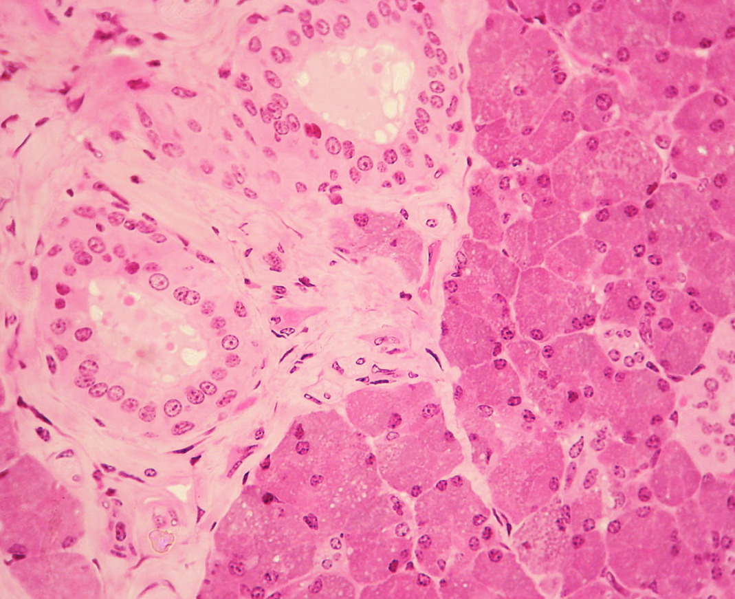
00:01 I want to now briefly summarize the salivary glands. On the left-hand side is a diagram illustrating the components of the salivary glands. You have four processes in the diagram and they illustrate various aspects of the structure of the secretory portions of a salivary gland, and then another structure illustrating the duct portion leading from those secretory units, and they're labelled various names that I will refer to. Each secretory unit of the salivary glands are called acini. And you can see in the diagram, and also, I will show you in the moment on the images of the sections through these salivary glands on the right hand side, the acini, the little berries or grape-shaped collections of cells that secrete the components of the salivary gland. It can be classified as either mucus or serous. And each of these acini then liberates their products in the central space or the luminal space of the acinus. And those products then pass through a ductus system that carries the secretion products to a large excretory duct, and therefore, into the oral cavity. That duct system begins with the intercalated duct right next to the acini. And that intercalated duct then passes to the striated duct. And those ducts are surrounded as of the acini with myoepithelial cells that help liberate the secretion product into the lumen from the secretory cells, and then move the secretion products along the duct system, because they contract all those myoepithelial cells. 02:08 Down the bottom on the diagram, it just illustrates the general structures inside the different secretory cells. And because of the different contents, these salivary glands stain very differently under some circumstances.If you now focus your attention to right hand three histological sections, I will point out the difference in the different salivary glands. The top section is the parotid gland. 02:40 When you look very carefully at the cells, they are serous secreting, which means they secrete a watery proteinaceous type product. And these cells, you can often see stain very pink. And what you're staining there are the granules, the secretory granules containing the salivary enzymes in the apex stored in the apex of the cells ready to be liberated. The rest of the cell is often stained basophilic, a bluey tinge, because that reflects the enormous factory for making the proteins in these granules, the rough or granular endoplasmic reticulum. On the next section is a sublingual gland. That is basically mucus secreting. All the cells there are a clear bluey pale stain. And most of the time in these glands, the mucus product is not preserved during processing for normal H&E sectioning and staining. 03:52 So a lot of the contents of the cell, the secretory contents, leak out. And so the cell appears to be rather vacant or empty. And so you see them rather very clear stained. And you?ll see more of those mucus-secreting cells in other organ systems I described in this histology course, where sometimes you just see whole clear cytoplasm. 04:15 And then at the bottom section is the submandibular gland. This gland is mixed. 04:22 It has got both serous components and it has got both mucus components. So that enables you, when you look at histological sections of all the salivary glands, to distinguish between parotid, sublingual, and submandibular. 04:43 The parotid is serous, the sublingual is mucus predominantly, and the submandibular is mixed. 04:53 You know, when I was a student, I could never remember that. I still can't really, but I have a little trick that helps me remember which one is serous, which one is mixed, and which one is basically mucus. And this may sound a bit funny but the parotid gland is the most superior of the salivary glands, S for superior, S for serous. And the submandibular gland has got an M in it, M for mixed, M for submandibular. I know that sounds funny, but it's the only way I can remember. And I suggest you try and think of another way to remember it or you can use my way. Before we move on, I want you to go back and look at the left-hand diagram, and I want to explain just something about the intercalated duct and the striated duct. 05:58 The intercalated ducts and the striated ducts are just not conduits, at least in serous secreting glands, because in serous secreting glands, they modify the secretion product. So they have a very important role in determining the final constituents of these salivary glands. So in serous secreting glands like the parotid, and we'll see later on when we look at the pancreas in another lecture, these intercalated ducts, and these striated ducts are quite prominent and reasonably easy to identify. But they're very hard to see in the submandibular gland, and the mucus-secreting components are very, very short. 06:48 The mucus components of the submandibular gland don't have very prominent intercalated ducts or striated ducts, whereas, the serous components will. 06:57 So often, they are very difficult to find in the submandibular gland. In the sublingual, you won't find it at all really because they are so short because they don't have any role in modifying the secretion product.The term striated duct refers to the fact that when you see these ducts, they have striations on the basal borders of the cuboidal epithelium that forms a duct, and this reflects very significant basal foldings of the basal cell membrane, and also lots of mitochondria. And this provides a much greater surface for the transport proteins and transport channels to move different products across that surface, and the mitochondria there to of course provide the energy for the active transport that's required. That's why they're called striated ducts.
About the Lecture
The lecture Salivary Glands by Geoffrey Meyer, PhD is from the course Gastrointestinal Histology.
Included Quiz Questions
The basophilic areas in the acinar cells of the parotid glands are a result of staining of the ....?
- Ribosomes
- Glycoproteins
- Mucigen granules
- Lysosomes
- None of the other answer options
Which of the following salivary glands is INCORRECTLY matched with the corresponding type of secretion?
- Sublingual - mainly serous fluid
- Submandibuar - mixture of both serous fluid and mucus
- Minor salivary glands - mainly mucus
- Parotid gland - mainly serous fluid
Which of the following salivary glands produce fluid that is mainly mucus?
- Sublingual glands
- Submandibular glands
- Parotid glands
- All of the salivary glands produce the same proportion of mucus secretions
Where are the myoepithelial cells found?
- In the glandular epithelium beneath the luminal cells
- In the mucosa above the basement membrane
- In the mucosa below the basement membrane
- In the glandular epithelium beneath the basement membrane
A glandular duct which is lined by simple cuboidal epithelium that contains numerous mitochondria is most likely which of the following types of ducts?
- Striated duct
- Intercalated duct
- Lobar duct
- Interlobular duct
Which of the following is INCORRECT?
- An intralobular duct is the portion of an endocrine gland inside a lobule.
- There are two types of salivary gland secretions - serous and mucous.
- Myoepithelial cells in ducts almost completely separate ductal epithelial cells from the basement membrane.
- Myoepithelial cells have contractile functions.
- Myoepithelial cells expel secretions from the lumen of secretory units.
Which ONE of the following is INCORRECT?
- The sublingual glands contain prominent intercalated and striated ducts.
- Intercalated and striated ducts help to modify salivary fluid.
- Striated ducts contain abundant mitochondria.
- The parotid gland and the pancreas contain prominent intercalated ducts.
- The parotid gland secretes mainly serous fluid.
Author of lecture Salivary Glands

Geoffrey Meyer, PhD
Customer reviews
5,0 of 5 stars
| 5 Stars |
|
1 |
| 4 Stars |
|
0 |
| 3 Stars |
|
0 |
| 2 Stars |
|
0 |
| 1 Star |
|
0 |
Very clear explanation and quick review after the topic explanation. Geoffrey also exposes tricks for remembering key items. I admire very much this professor.
