Playlist
Show Playlist
Hide Playlist
Acute Pancreatitis: Definition, Etiology and Diagnosis

-
Slides Pancreas and Biliary Tract.pdf
-
Download Lecture Overview
00:02
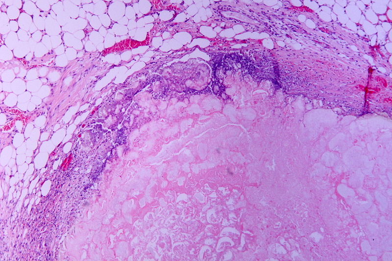
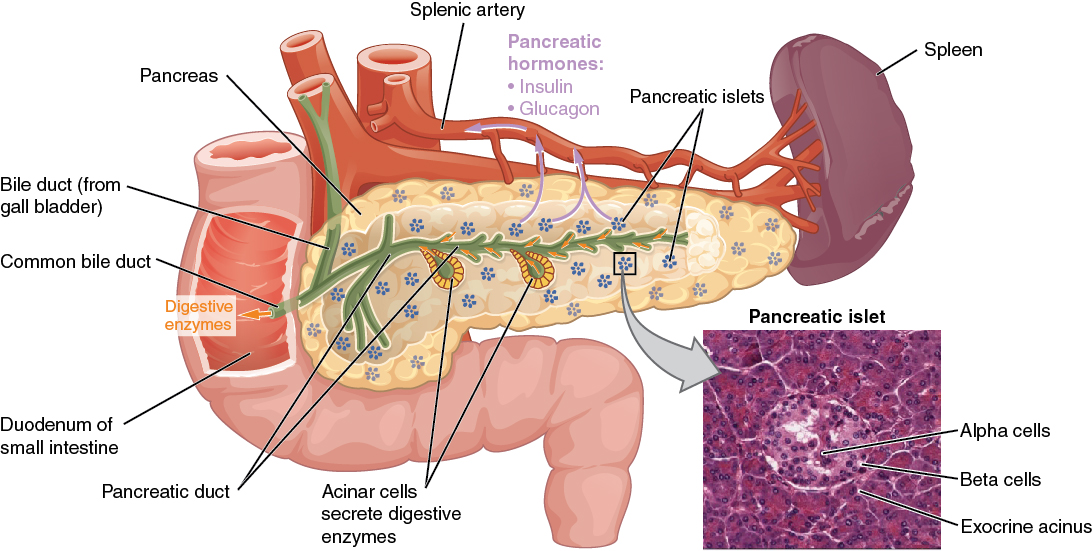
Acute pancreatitis, by definition,
everything in your pancreas is
going absolutely crazy. In your head, you
should be thinking about the pancreas
and divide it into two physiologic organs.
And by that, I mean you divide it into
exocrine pancreas and you divide
it into endocrine pancreas.
00:23
Initially, when there's damage that
has taken place to the pancreas,
all these enzymes that are being released
and, in your head, you should also
be thinking about, "Whoa, those enzymes
from the pancreas that are responsible for
protein digestion include trypsin,
carboxypeptidase, endopeptidase
and so forth, trypsin being
the most potent.
00:43
If you're dealing with the enzymatic
or exocrine pancreas and you wish
to metabolize your lipid and then
that discussion takes you to lipase.
00:51
In any case, that's a lot of enzymes
within the pancreas, is it not?
And all these enzymes are being
released locally, you might have
utmost enzymatic destruction resulting
in a term called autodigestion.
01:07
Important etiologies that we'll take
a look at: alcohol being quite common.
01:11
This pain that the patient's
feeling is not only epigastric,
but then also radiating to the back.
When the time is right, I will then
then give you a list of differentials
quickly to have you differentiate
between different types of epigastric pain.
01:25
Gallstones could also be a
possible cause of pancreatitis
and we'll talk about that in greater detail
and it's a nice little story.
01:32
In other words, you had a gallstone
that was in my gallbladder,
ended up in my biliary tree and
eventually makes its way to the
the second part of the duodenum in
which it may then cause what's known
as compression atrophy.
01:45
Other causes also, hypercalcemia,
may cause damage to the pancreas.
01:49
And, for example, if you have a patient
that has a type 4 type of hyperlipidemia
or even worse case, a type 1
hyperlipidemia.
01:58
And if that lipoprotein lipase
is not present, "Up goes my
chylomicron. Oh my goodness,
my triglyceride levels are ridiculously
high! Thousands."
The first organ to be damaged
here would be the pancreas.
02:12
Drugs, I'll give you a list of drugs
here that cause acute pancreatitis.
02:16
We have AZA or azathioprine.
We have pentamidine, and we have
hydrochlorothiazide, being important drugs.
There's more to come.
02:25
Infections such as mumps. With mumps,
even though it might be rare,
you want to keep this mind because
it may then cause parotitis, orchitis.
02:39
It might be in my best interest
not to grab my testicles.
02:42
We have coxsackievirus and we
also have parasites that may
all cause acute pancreatitis.
02:51
Embryologic issue that you might have
called pancreatic ductus divisum
and remember with the proper embryologic
development you have though,
the ventral head and the dorsal head,
and all these may then result in
an abnormal type of division
called a ductus divisum
or there might be inherited
autosomal dominant disorder
resulting in acute pancreatitis.
03:16
Interesting enough, you were performing
a procedure in which you were trying to
identify the pathology in your bile duct.
In the process, you actually ended up
causing damage to the pancreas, that's
a risk factor when implementing endoscopic
retrograde cholangiopancreatography.
That post exam, you might actually bring
about damage to the pancreas or just
straight up blunt trauma to the abdomen.
03:48
Important etiologies for acute
pancreatitis: Vascular,
we have ischemia or perhaps
vasculitis resulting in acute pancreatitis.
03:58
Miscellaneous include, well, importantly,
cystic fibrosis. So imagine a child that
has extremely viscous fluid
within his or her ducts.
04:12
Most likely a child of Caucasian
decent and with that viscous
type of fluid within the ducts,
not only could it result in
what's known as your bronchiectasis and
eventually result in pneumonia
because you have increased thickness
there, but they might have
increased thickness within the
pancreatic ducts. So therefore,
anatomy, the pancreatic duct is moving
towards the second part of the duodenum.
04:34
The fluid is moving in that direction.
04:36
If you end up having all those
viscous fluid within your duct,
and imagine now, that you may then
cause backup congestion and
compression and eventual acute
pancreatitis, an important point,
or the two types of ulcers that
we discussed earlier.
04:53
If your patient is giving you the
following symptoms initially:
"Hey, doc. I have this pain at midnight,
late at night when I'm asleep.
05:05
I have to go downstairs to the kitchen,
and I have to open up the fridge
and I end up having a pretty decent
amount of cake that we had for
yesterday's birthday and actually,
the pain then went away."
So initially, what I'm giving you here
are the symptoms of duodenal
peptic ulcer disease in which
the pain then has been relieved
by eating but then eventually what
may then happen, if not properly taken of
care of, is that this may then perforate.
And when it perforates, imagine
where you are. A duodenal peptic ulcer
disease, where are you? Think.
05:39
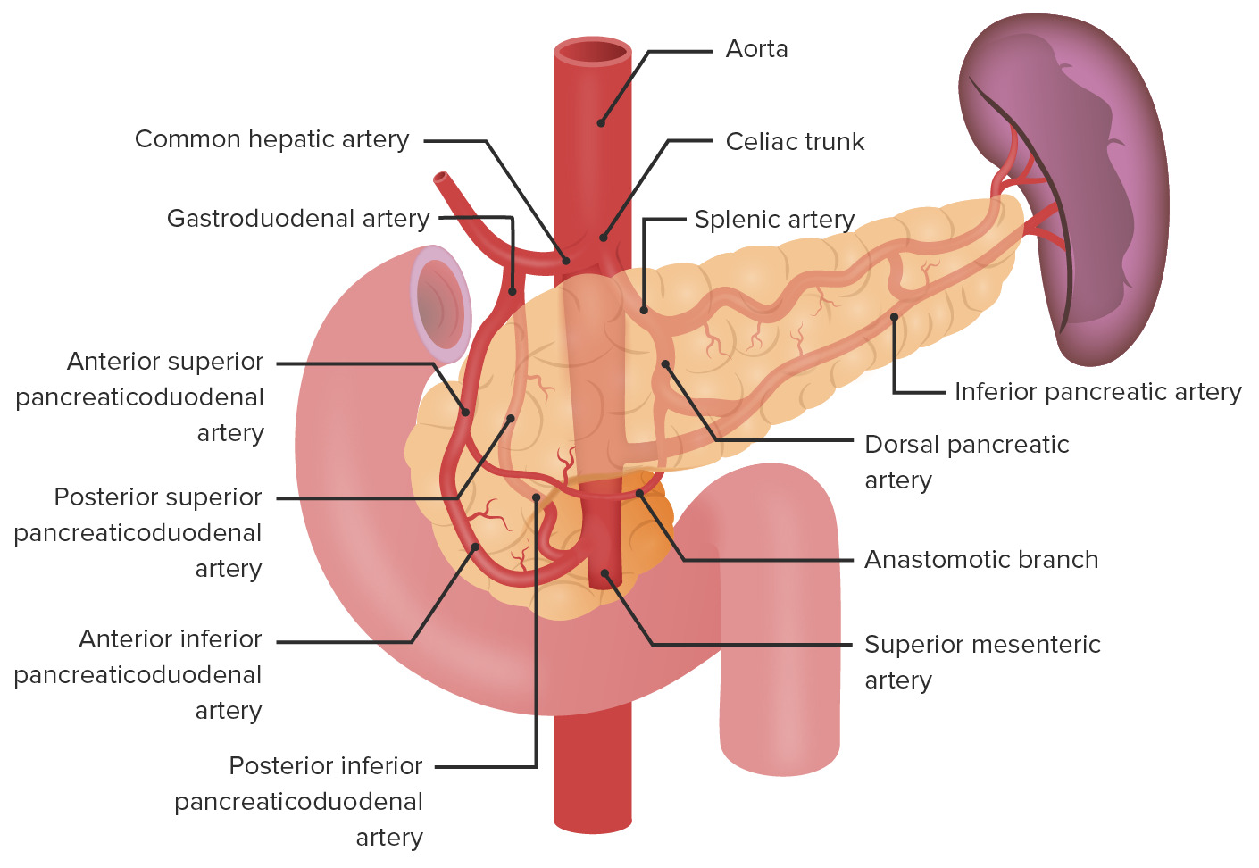
First part of the duodenum. If you were
to perforate through, you may then cause
damage to the gastric duodenal artery and,
in fact, then bring about damage
to the pancreas. Important. Miscellaneous?
Yes, but incredibly clinically important.
05:54
Signs and symptoms. As I told you,
I'll give you some important epigastric
pain issue. Here, with acute pancreatitis,
the patient is definitely feeling pain
in the abdomen, but then it radiates
to the back because of the
rectoperitoneal type of journey of the
head of the pancreas towards the tail,
meaning to say that it's going to be
up with the spleen, is it not?
If this gets damaged, then the patient
is also expressing pain in the back as well
I just gave you another epigastric pain
where the pain then went away after
consuming food and that's your
duodenal peptic ulcer disease
or you could have a patient with
epigastric pain and it and it does not
radiate to the back, in which the pain
gets worse immediately after eating
and that will be gastric type of
peptic ulcer disease.
06:41
Epigastric pain, their differentials
become important here.
06:43
Low-grade fever, nausea and vomiting
or signs and symptoms that you can expect,
but you'd be finding that with
any type of -itis, so obviously
giving you more information.
06:56
There might be tachycardia, orthostasis
or perhaps even ileus due to obstruction.
07:02
And a couple of important things here,
these might be rare in clinical discovery;
however, keep in mind because if you
find something like this in a stem
of a question or you see a picture
of what's known as Cullen's sign.
07:14
In other words, there'll be
hemorrhage around the umbilicus. C-U-U-M.
07:22
Cullen is bleeding around
the umbilicus or there might be
hemorrhage in the flanks and the flanks
will be the sides of your body,
out back towards the hips and
basically where you find you kidneys.
07:36
Are we clear? And around there,
if you ended up finding hemorrhage,
we then call this more or less
hemoperitoneum or rectoperotineal
hemorrhage. This will be referred
To as being your Grey-Turner's sign.
07:48
Rare as it may be, keep this in mind
because remember on your boards,
they have to give you specific
information so that you're moving
in a certain direction and you're
choosing one answer choice
over the other with almost utmost
confidence. Now that's it.
08:03
Obviously, there's always going
to be a little bit of doubt, trust me.
08:07
Always, but that's okay though
as long as you choose the most
educated answer then you'll be in
good shape, you move on.
08:17
Lab testing for acute pancreatitis
specifically, keep in mind that amylase
and lipase may be found, but which
one's more specific for acute pancreatitis?
In fact, it's lipase.
08:27
One thing interesting once again
that we'll see is that the enzymes
that you would find to be elevated
in acute pancreatitis may not correlate
with the severity of the disease.
We had this very discussion when we did
liver disease and the transaminases.
Even though the transaminases being
elevated would tell you that the
patient most likely has liver disease.
08:47
It does not correlate with the severity.
Same concept here as well.
08:54
Serum liver function test: if elevated,
may then suggest gallstone pancreatitis.
08:58
We will talk about gallstones
in great detail. And with gallstones,
at least think about this before
we actually get to the details.
09:07
You can have a stone that
begins in the gallbladder.
09:10
Let's say it's cholesterol stone or
a pigment stone if there is bulirubin.
09:14
That stone may then escape
from the cystic duct and
you are then moving throughout
the biliary tree and as
then moves through the biliary tree,
which is then technically called your
your choledocholithiasis, understand
that you might have compression issues
and also injury that might be
taking place to adjacent organ,
including the liver and then also
including the pancreas.
09:39
We'll talk more later, it's important.
09:41
Imaging studies. You will do an abdominal
and, stop -- chest x-ray.
09:46
What' Now, understand what's going on here,
don't just memorize this.
09:50
Where is the pancreas located?
Right, in the abdomen. Okay.
09:54
Then why the chest? Is there such a thing
in which you have a hernia
in which the pancreas comes up
in the thorax? Are you kidding me?
I suppose anything is possible, but listen,
this is why you're doing a chest x-ray.
10:06
This pancreas has been damaged, okay?
You do an abdominal x-ray and
you might find 'a sentinel loop.'
Stop. The pleural effusion and ARDS
have nothing to do with the abdomen
and why is it associated with pancreatitis?
Enzymes. What does ARDS mean?
Acute respiratory disease syndrome.
It means that the alveoli
had been destroyed. There's every
possibility that enzymes being released
from the pancreas into circulation
may then cause damage to the lung
extensively to the point where
the pleural cavity may then develop fluid
and the alveoli may then
perish. Is that clear?
Therefore, what you're looking
for in a chest x-ray would be atelectasis.
10:56
CT: Useful for evaluation of
complications, normal in about
30% of your patients, however.
11:03
ERCP: Reserved for therapeutic
interventions, and by that we mean that -
Remember, ERCP itself, even though you're
trying to explore what's going on with
with pancreas and what's causing
damage, in the process you might
actually cause damage to the pancreas,
so be careful with ERCP.
11:21
So it could be diagnostic, but then also
be an etiology of lesion.
11:27
MRCP, obviously playing a much
greater role. We have MRI,
is what the MR stands for,
and it's non-ivasive,
increasingly replacing the ERCP
for obvious purposes.
About the Lecture
The lecture Acute Pancreatitis: Definition, Etiology and Diagnosis by Carlo Raj, MD is from the course Pancreatic and Biliary Tract Diseases: Basic Principles with Carlo Raj.
Included Quiz Questions
Which of the following is NOT a common risk factor for acute pancreatitis?
- Smoking
- Alcohol
- Hypertriglyceridemia
- Gallstones
- Hypercalcemia
For which of the following genetic diseases is acute pancreatitis a common complication?
- Cystic fibrosis
- Hemochromatosis
- Wilson's disease
- Phenylketonuria
- IgA deficiency syndrome
A 20-year-old man occasionally consumes alcohol and has a serum triglyceride level of 1,000 mg/dL. He was diagnosed with acute pancreatitis, and his cousin was treated for acute pancreatitis and alcoholic cirrhosis 1 month ago. What is the probable cause of this patient's acute pancreatitis?
- Hypertriglyceridemia
- Alcohol
- Drugs
- Hereditary pancreatitis
- Gallstones
A patient who presents with congestive cardiac failure developed muscle weakness after treatment. After a few days, she presented with acute pancreatitis. Which drug is the probable cause of the acute pancreatitis?
- Hydrochlorothiazide
- Spironolactone
- Digoxin
- Propranolol
- Furosemide
A 10-year-old boy presents with swelling and pain in the parotid regions and testicles. He began to develop epigastric pain. What is the MOST likely cause of epigastric pain?
- Acute pancreatitis
- Acute cholecystitis
- Acute hepatitis
- Acute appendicitis
- Acute peptic ulcer disease
Which procedure can cause acute pancreatitis as a complication?
- ERCP
- MRCP
- Upper GI endoscopy
- Ultrasound-guided FNAC
- X-ray of the abdomen
What is the sign on clinical examination that shows discoloration around the umbilicus?
- Cullen's sign
- Cushing's sign
- Caput medusa
- Kosher's sign
- Sitkovsky's sign
Which of the following is NOT associated with Grey-Turner's sign?
- Chronic pancreatitis
- Acute pancreatitis
- Blunt trauma to abdomen
- Retroperitoneal hemorrhage
- Ruptured ectopic pregnancy
At which of the following sites can a gallstone become impacted, leading to acute pancreatitis?
- Sphincter of Oddi
- Right hepatic duct
- Left hepatic duct
- Cystic duct
- Neck of the gallbladder
An increase in which of the following enzymes indicates acute pancreatitis?
- Pancreatic amylase
- Salivary amylase
- Pepsin
- Gamma-glutamyl transferase
- AST
Author of lecture Acute Pancreatitis: Definition, Etiology and Diagnosis

Carlo Raj, MD
Customer reviews
4,7 of 5 stars
| 5 Stars |
|
2 |
| 4 Stars |
|
1 |
| 3 Stars |
|
0 |
| 2 Stars |
|
0 |
| 1 Star |
|
0 |
Such personality! Loved your lecture Dr. Raj! Keep up the good work!
the lession is very insteresting, but i think i could be explained a little better, showing and explaining an image of CXR of atelectasis, sentinel loop AXR. Also it coulde be better if hidroclorotiazied mecanism way of producing muscle weakness is explained after answering the question of the quizz, and the indications of the ERCP explained. Thanks
The review was easy to understand with clear explanation. I also appreciated the test sample after the lecture. It tested my knowledge regarding the subject matter thank you
