Playlist
Show Playlist
Hide Playlist
Golgi Apparatus

-
Basic Histology 02.pdf
-
Download Lecture Overview
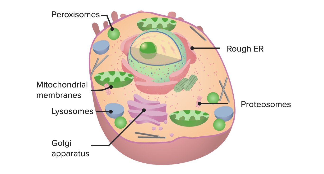
00:01 Let's now move on to another very important organelle in the cell and that is the Golgi apparatus or Golgi complex. 00:11 Golgi or Golgi, it's pronounced either way. 00:16 Have a look at the image or the diagram and it shows you the structural appearance of the Golgi apparatus. 00:27 They're really parallel stacks of tubules and also little vesicles. 00:36 They really are highly associated with the nucleus and also the rough endoplasmic reticulum. 00:46 You can see the nucleus and the rough endoplasmic reticulum in a paler version in the diagram on the right-hand side and they have what we call a cis face or an entry face and a trans face or an exit face. 01:05 And the reason for that is that one of the jobs the Golgi apparatus does, and I'll mention that in a moment, is that it accepts proteins and various components synthesized by the endoplasmic reticulum. 01:21 It accepts those products at the cis face or the entry face and then those products pass through the Golgi complex where they're sorted and packaged and then they leave at the exit face or the trans face. 01:42 I always get mixed up sometimes at what is a cis and what is a trans face, and I therefore think that while trans means transport, transport is what the Golgi apparatus does at its final role and therefore, that's at the actual exit or the trans face of the Golgi complex, trans or transport away from the products being sorted and modified, that occurs at the last station, if you like, of the Golgi complex which is the trans face as opposed to the cis face which is the entry point. 02:27 And besides sorting and packaging, the Golgi apparatus is involved in making carbohydrates, it modifies proteins made by the endoplasmic reticulum, and as I've already mentioned, it sorts and packages them for secretion or transport within the cell in these little secretory granules or vesicles you see in the diagram. 02:55 Here's a good example of the function of both the Golgi apparatus and the endoplasmic reticulum. 03:06 Here, you have protein synthesized by the endoplasmic reticulum and they're packaged into a transport vesicle. 03:15 That transport vesicle then enters the cis face of the Golgi complex and the components or the products pass through the Golgi complex where they're sorted, modified, and then repackaged into a secretory vesicle which then passes to the cell membrane, and the secretory vesicle itself fuses with the cell membrane and releases the secretory product. 03:46 That process is called exocytosis. 03:52 Now, you can even see Golgi apparatus in the light microscope pictures. Very difficult. 04:01 In this image, if you scan around and I know it's going to be very hard for you to find this, but on the left-hand side it says, "Look for a clear area in the cell cytoplasm adjacent to the nucleus." If you run your eyes along the first part of that sentence, "look for a clear area in", you follow your eyes across onto the image, you will see a cluster of 1, 2, 3, 4, 5... about 7 or 8 cells just left of the middle of the image. 04:36 Well, if you look carefully at the top nucleus of one of those cells, the one that's a little bit on its own, you can just make out a very, very pale area near that nucleus. That represents the Golgi apparatus. 04:53 I know that's a very hard thing to do, but when you study more histology, you become more experienced at doing this. 05:01 Try again. Again, look for a clear area in, follow your line of sight across to the middle of the image, you'll see a cluster of about 6 or 8 cells, the one on the far left of that group again has a very clear area right near the nucleus that represents the Golgi apparatus in the light microscope. 05:26 Very difficult to see though, I must admit. 05:30 A bit easier when you look at the electron micrograph of the Golgi apparatus shown here. 05:35 You can see, it's the stacks of membranes you see right in the center of this image. 05:42 Above the stacks of membranes, you can just make out some profiles of mitochondria, and the darker-stained circular structures are going to be lysosomes, which I'm going to speak to you about in a moment.
About the Lecture
The lecture Golgi Apparatus by Geoffrey Meyer, PhD is from the course The Mammalian Cell.
Included Quiz Questions
What is the main function of the Golgi apparatus?
- Packaging and transfer of proteins
- Protein synthesis
- Lipid synthesis
- Enzyme degradation
- Synthesis of adenosine triphosphate
Author of lecture Golgi Apparatus

Geoffrey Meyer, PhD
Customer reviews
4,5 of 5 stars
| 5 Stars |
|
1 |
| 4 Stars |
|
1 |
| 3 Stars |
|
0 |
| 2 Stars |
|
0 |
| 1 Star |
|
0 |
Amazing and I learned more from his videos then in class.
I chosed to give it 4 stars, because I loved the way the lecture was eplained. I am not a native english speaker and therefor prefered the rather slow speed of speach. unfortunatelly there have not been any Control questions and that is why I only rated this lecture with 4 out of 5 stars.
