Playlist
Show Playlist
Hide Playlist
Cytoplasmic Inclusions, Cytoskeleton and Interphase of the Cell Cycle

-
Basic Histology 02.pdf
-
Download Lecture Overview
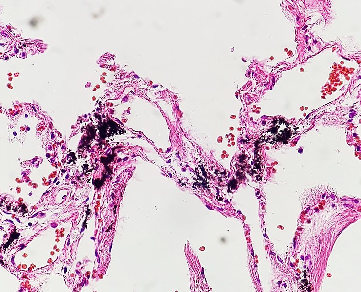
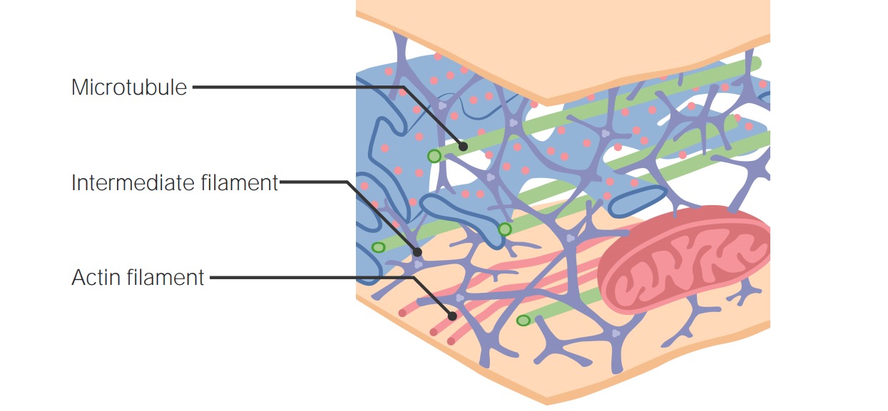
00:01 Now, let me concentrate on cytoplasmic inclusions. 00:06 They're the nonliving substances of the cell and they're not bound by a membrane and they include lipid droplets, zymogen granules, and glycogen granules that we see in the digestive glands and also the glycogen granules we see in the liver listed here, protein crystalloids which I'm not going to include because although they're mentioned here, their function is essentially unknown in most parts or in most tissues of the body. 00:44 And then finally, we have pigment granules you see in some cells. 00:48 And then another inclusion are going to be secretory granules, but they're membrane-bound. 00:56 Here is a section through the adrenal cortex and in the middle, you can see a very large cell with a nucleus and it's full of little round, circular, white structures. 01:12 They're lipid, lipid droplets. They dominate the cell and that's because these particular cells, as all cells do in other parts of the body, not just the adrenal cortex—you find them in the ovary and the testes, for instance. 01:28 These cells are making steroid and the lipid droplets are cholesterol stores, etc. 01:36 They're the precursors for all the production of the steroid hormone that the cell is producing. 01:43 They appear clear in the cytoplasm because being of a lipid component. 01:50 That lipid component is lost during the processing of tissue and therefore, they just appear as a ghostly white structure within the cell. 02:02 Here on the left-hand side, you can see a chain or a network of cells in the liver. 02:08 They're all hepatocytes. 02:10 You don't see much detail of the cell. You can occasionally see a small pale-blue circular nucleus, but the pink-stained component is glycogen. 02:24 And you can see on the right-hand side a very high magnification view at the electron microscope level. 02:31 You can see the glycogen appearing like soot scattered throughout the cell. 02:37 Well, that glycogen is stored in these hepatocytes and is the energy source we use, or one of the energy sources we use in our bodies. 02:48 You don't normally see it in normal histological sections. You need a special stain to see that glycogen, and I'll talk about staining and what stains indicate in another lecture in this series. 03:05 Here is a rather high magnification picture of the skin, and you can see a curved line of cells and nuclei, and you see this brown pigment, that brown pigment we have in all our skin cells and that pigment is melanin. 03:27 And melanin protects our skin, the very basal cell layers of our skin that protects those cells from any damage from ultraviolet light. 03:40 The very basal cells of our skin are dividing constantly to give rise to layers of our skin that form the remaining epidermis or our top layer of skin. 03:54 And as cells are formed in this basal layer pass up through the different layers of the skin surface, they finally are transformed into keratin which you see on the very surface of this particular slide. 04:09 That keratin is our waterproofing agent, so this melanin protects those basal dividing cells from any DNA damage that may be caused by excessive ultraviolet light. 04:24 And here you see a diagram of a cell called the melanocyte sitting in the basal layer of the epidermis or the cells that make up the layer of the skin surface. 04:37 Those melanocytes are very specialized cells that make the melanin in response to sunlight, and then they transfer that melanin into the neighboring skin cells, particularly the layer just above the basal layer, the stratum spinosum, and those cells in the stratum spinosum actually ingest the melanin and the melanin aggregates around the top surface of the nucleus to protect it from the sunlight or ultraviolet light coming through. 05:15 So melanin in this particular cell population is a cell inclusion that's extremely important. 05:24 There is also neuromelanin which is a similar product that we have in the retina of our eye. 05:30 If you look at this diagram at the very top, you see the choroid layer with the pigment epithelium is the very external layer of the retina shown here in this particular diagram. 05:43 That pigment epithelium, as we'll learn about when you study the eye in more detail, has a number of essential functions in the retina, and again, an example of a cytoplasmic inclusion. 05:59 Some cells such as neurons, skeletal muscle cells and cardiac muscle cells, cells that are fully differentiated, they don't undergo division. 06:12 When they break down components that I spoke about earlier under the action of these lysosomes, the breakdown products remain inside the cell as residual bodies or age pigment, and it's got a yellowy-orange stain to it or color to it. 06:29 We call it lipofuscin, and there is some in these particular cells, but I'm not going to ask you to see them or try and point them out here. 06:40 Instead, there will be a label added or a pointer added to this image to show you lipofuscin after you look at it just for a moment or so. 06:52 Now, you can see a similar pigment to lipofuscin in these cells here. It's called hemosiderin. 06:59 Hemosiderin is the residual components of the breakdown of hemoglobin. 07:07 Red blood cells only live for about 120 days and when they pass through the spleen, the old ones can't squeeze through all the network of barriers really the spleen puts up to test whether these red bloods can squeeze through. 07:27 The old ones can't so they get caught in the network of fibers or networks of reticular fibers that are set up in the spleen, and macrophages come along and break them down. 07:39 They break them down and reuse part of the components of hemoglobin. 07:43 The body is very much into recycling, but the residual amount you see here in the cytoplasm of macrophages within the spleen, that orangey-browny component you can see in this particular image, hemosiderin. 08:03 Secretory granules are membrane-bound and they're produced by cells, particularly protein-secreting cells, particularly all our glands that secrete digestive enzymes, saliva, etc. 08:18 And in this image, you can see in these groups of cells very pink-staining, tiny little granules. 08:27 Forget about the right-hand side where there's a lot of white-staining cellular matter. 08:32 That's a different part of this particular tissue, the pancreas. 08:36 Focus on the central area where you see a cluster of cells, and they have very fine pink granules within them. 08:45 They're secretory granules and they will pass to the cell surface and then out through the lumen and then going through a duct system to the part of the body where they're utilized. 08:59 And again more clear on the right-hand side you see lots of secretory granules in this particular cell. 09:05 It doesn't matter what it is. 09:07 It really is just to show you the predominance of these cytoplasmic inclusions being secretory granules which are membrane-bound. 09:18 On the left-hand side, you see them in a much lower power in a salivary gland just appearing as little tiny dark dots within the cell cytoplasm. 09:33 Lastly, let me just focus on two points. One is the cytoskeleton. 09:39 And again, I'm not going to go into great details of the cytoskeleton which is beautifully illustrated in the image on the right. 09:48 The image on the right depicts all the different components of the cell cytoplasm which is really microfilaments, microtubules and intermediate filaments stained in different colors there, and I don't really want you to worry about which is which in this particular slide. 10:09 Focus more on how extensive it is in the cell and as I might have mentioned earlier, the cytoskeleton supports the cell membrane and it's also involved with moving organelles around the cell and also supporting all those organelles within the cell. 10:28 When we look at epithelia, we will see that the cytoskeleton is also involved in reinforcing projection from the surface of epithelia such as microvilli, cilia and also flagella in the sperm of the male gamete. 10:52 Finally, I just want to briefly mention the word interface that I mentioned in the very first part of this lecture when we spoke about the nucleus. 11:04 Interphase is the part of the cell cycle where the nucleus is not undergoing structural change for cell division that occurs during mitosis which you see depicted in this diagram. 11:19 And I don't want to go into the process of mitosis here which is depicted on the right-hand side prophase, metaphase, anaphase and telophase. I'll leave that to your study of genetics or other parts of your cell biology. 11:36 But finally, let me just bring your attention to again this interphase part of the cell cycle shown on the left-hand side of the diagram, and the G1 phase is the most different in length. 11:54 It's the most prolonged part of the cell cycle. 11:57 You're going to see nuclei in all your cell populations in your histology course that are in either G1, the S or the G2 phase but structurally, the nuclei will look very similar in all those three phases. 12:14 Lastly, some cells leave the cell cycle. 12:20 They go into the G0 phase, meaning outside. They're outside the cell cycle because they no longer want to divide, and those cells are the neurons in our body, in our spinal cord and brain, cardiac muscle cells, and skeletal muscle cells, and those cells cannot be replaced because they've left the cell cycle, so when neurons die, when cardiac muscle dies, then that's the end of those particular cells. 12:55 Skeletal muscle, on the other hand, has the ability to repair itself if it's damaged, but they are what we call in a fully differentiated state. 13:05 They no longer have the ability to divide and grow and further differentiate.
About the Lecture
The lecture Cytoplasmic Inclusions, Cytoskeleton and Interphase of the Cell Cycle by Geoffrey Meyer, PhD is from the course The Mammalian Cell. It contains the following chapters:
- Cytoplasmic Inclusions
- Cytoskeleton
Included Quiz Questions
Most fully differentiated neuronal cells reside in which of the following cell cycle phases?
- G0
- G1
- G2
- S
Author of lecture Cytoplasmic Inclusions, Cytoskeleton and Interphase of the Cell Cycle

Geoffrey Meyer, PhD
Customer reviews
5,0 of 5 stars
| 5 Stars |
|
5 |
| 4 Stars |
|
0 |
| 3 Stars |
|
0 |
| 2 Stars |
|
0 |
| 1 Star |
|
0 |
