Playlist
Show Playlist
Hide Playlist
Endocrine Pancreas

-
Slides 05 Human Organ Systems Meyer.pdf
-
Download Lecture Overview
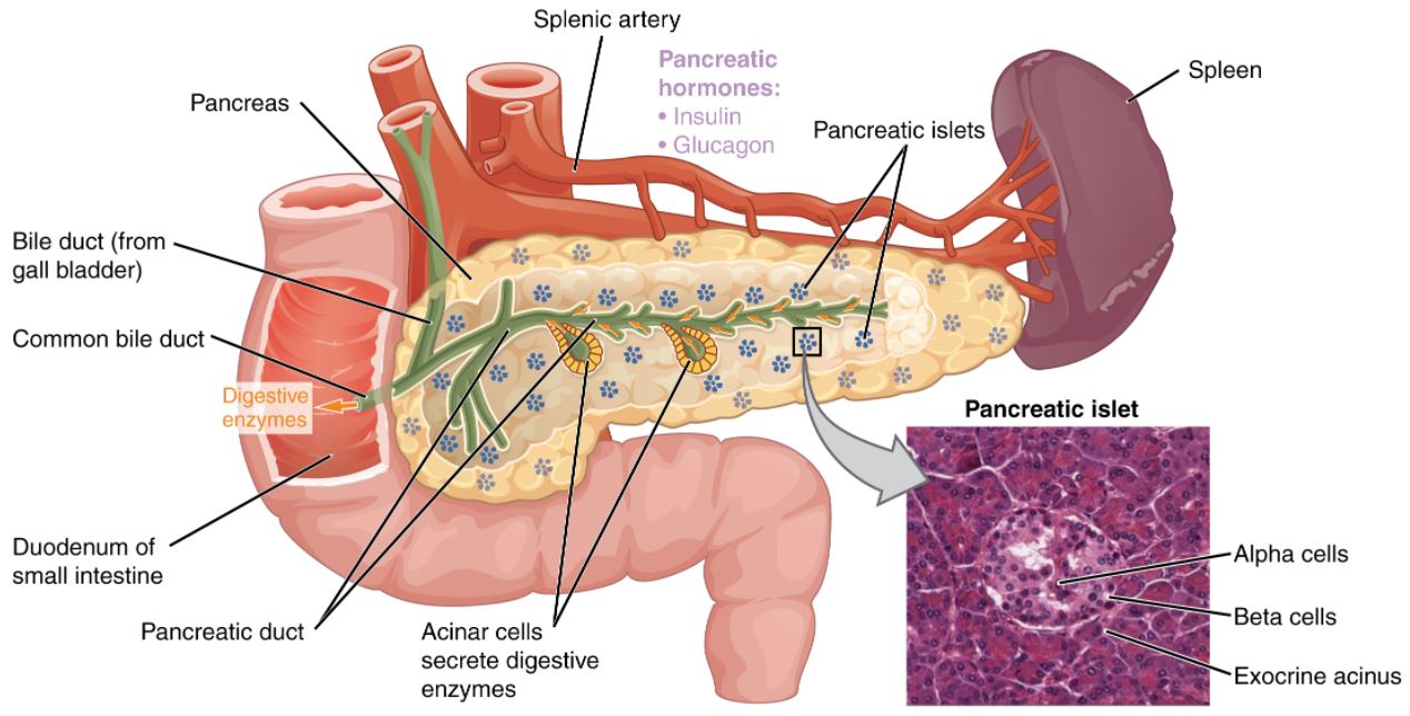
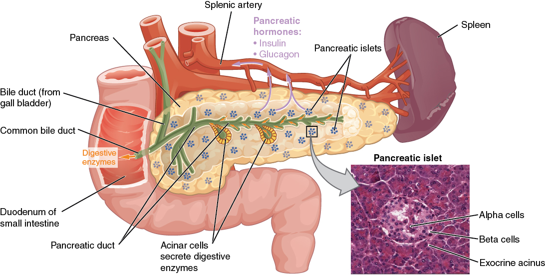
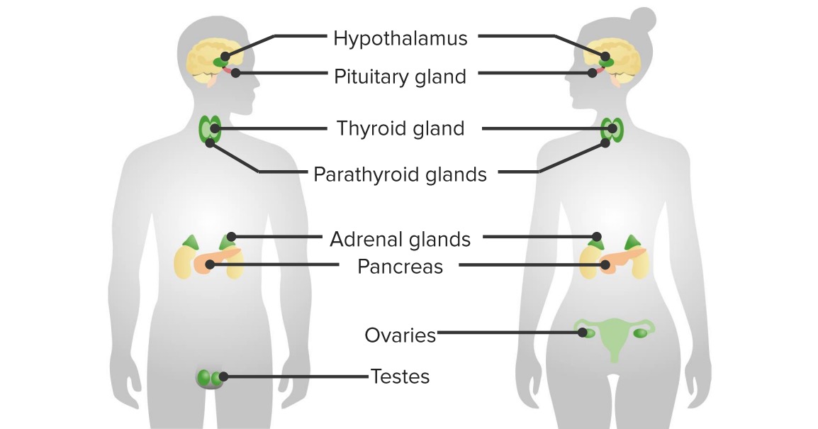
00:01 So it’s a specialized vessel, having a specialized function. Well, last of all, let’s look at pancreas. When you look at the pancreas, it has two different components. It’s an exocrine gland that in fact, there’s 90% or even more of its histological components, an exocrine gland that secretes pancreatic enzymes. And those enzymes go into a duct system, and the duct then goes and deposits these secretory products into the duodenum to help with the digestion of the food in which we ingest. Well, the other component is the endocrine component. It’s the component that occupies a very very small percentage. 00:45 And you can see in sections of the pancreas as you see here on the left-hand side, a section through the pancreas showing one of these endocrine components that appears the pale staining little ball of cells. The darker staining on the periphery are the exocrine components, the serous secreting exocrine acini. But focus on the central paler stained component. 01:14 That is the endocrine component. It’s very vascular, even though you don’t appreciate here for reasons I mentioned earlier, the vessels that capillaries tend to collapse down, but they are fenestrated capillaries again. Well, these endocrine components are called islets of langerhan, or more recently, we refer to them as pancreatic islets. And they can be of different sizes. Some can occupy only a hundred cells. Some can be very, very large. So there’s this variety and structure in the size of these islets just like I explained there was a variety in the diameter and size of the thyroid follicles we looked at the very start of this lecture. On the right-hand side is another preparation used to try and distinguish the three major cells types in these islets. I’m not going to point them out here and try and distinguish them, but you can see that there are differences, there are different sorts of cells. Some are eosinophilic, some are basophilic and some are not so basophilic. 02:25 So if we look inside one of these pancreatic islets indicated in the left-hand side, and then look on the right-hand side and try and identify the cells, basically, you can class the cells as either A, B, or D cells or alpha, beta, and delta cells. And really, they refer to the hormones that they secrete. And in H&E sections as you see here, you cannot distinguish these real cell types. So I wouldn’t expect you to. But I’d like you to remember that the A cells secrete glucagon, which controls glucose levels that we’ll talk about at another time. The B cells by far occupy the most cell numbers in these pancreatic islets. 03:14 They secrete insulin. And the D cells secrete somatomedin and a couple of other components that we are not quite sure of their exact function yet. But certainly, the endocrinologist will talk to you or describe to you what exactly these B cells do secrete. But focus on the A and the B cells, those that secrete glucagon, and those that secrete insulin. Both those hormones have very, very important widespread functions throughout the body. In summary, it’s important that you understand the histological difference between each of these endocrine glands that I’ve described in this lecture. Know what sort of cells are in each of these four endocrine glands or tissues, know the hormonal products each of these glands secrete, and their general effects on their target organs. And know also a little bit about specialized components of each of these glands, such as the blood supply in the adrenal cortex, and how it affects the adrenal medulla. And also, the different way in which the thyroid gland operates. It secretes and stores its product and it reabsorbs it. So when you look at the structure and function of all these glands that I’ve described, you should be able to point out all their differences, structurally and functionally. But remember, together, collectively, they are very important components of the endocrine system. So thank you for listening to this lecture. I hope you now have a good understanding of these very, very important endocrine glands.
About the Lecture
The lecture Endocrine Pancreas by Geoffrey Meyer, PhD is from the course Endocrine Histology.
Included Quiz Questions
The beta cells of the pancreas secrete which of the following hormones?
- Insulin
- Glucagon
- Somatostatin
- Adrenaline
- Cortisol
The delta cells of the pancreas secrete which of the following hormones?
- Somatostatin
- Glucagon
- Insulin
- Cortisol
- Adrenaline
Which of the following statements regarding the pancreas is INCORRECT?
- The islet cells are a type of chromaffin cell.
- It is both an exocrine and endocrine gland.
- It contains both alpha and beta cells.
- Delta cells secrete somatostatin.
- Alpha cells secrete glucagon.
Author of lecture Endocrine Pancreas

Geoffrey Meyer, PhD
Customer reviews
5,0 of 5 stars
| 5 Stars |
|
2 |
| 4 Stars |
|
0 |
| 3 Stars |
|
0 |
| 2 Stars |
|
0 |
| 1 Star |
|
0 |
Well broken down. Thank you. I love that he takes the time to analyze the explanations.
This was so easy to grasp, that you so much!
